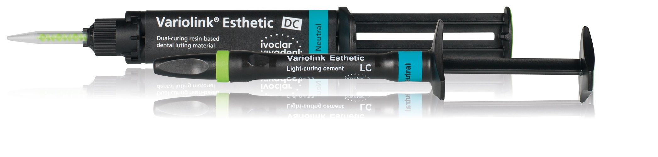
Variolink Esthetic
Variolink Esthetic is an esthetic light - and dual-curing luting composite for the permanent cementation of ceramic and composite restorations. The cement features excellent shade stability, lifelike fluorescence, and flexible consistency.
Ivoclar
800-533-6825 | ivoclar.com
Americans are living longer1 and many are remaining in the workforce well beyond retirement age.2,3 Individuals 60 years and older have retained more of their teeth than previous generations4 and, driven by an increased awareness of esthetics, tend to place great importance on retaining a youthful, healthy smile.5-8
A common problem for dentists when considering esthetic restorative options for the aging workforce is the loss of interdental papilla and the formation of maxillary and mandibular open gingival embrasures, or black triangles.9 Traditional restorative treatment solutions may fail to achieve ideal esthetics. However, adapting to the bone height in the interproximal area can offer an effective solution.
Many workers plan to remain in the workforce beyond age 70—or not retire at all.1 For those who plan to continue working, aging can be a competitive disadvantage for retaining their position.10 Esthetic dentistry is a key factor to combating aging and restoring a more youthful appearance for these patients.7
For esthetic dentists, treating these patients may pose a challenge. Due to years of restorative procedures in the hopes of maintaining their natural teeth, a host of patchwork dentistry may exist in the esthetic zone. These past treatments, in combination with the effects of aging, often result in the formation of open interdental embrasures in the maxillary and mandibular arches that are perceived as unesthetic by the patient and as a risk to good oral health by the dentist.11,12
Case Presentation
A female in her 60s was referred to the practice for restorative consultation and treatment. Her chief concern was the appearance of her smile, in particular the shape and color of teeth #7 and #10 as well as the black triangle between teeth #8 and #9. She was disappointed with failed attempts of previous dental work and was skeptical that any treatment plan would give her the smile she wanted. Her goal was to rejuvenate the natural appearance of her smile without the treatment being obvious to others.
At the initial consultation, a series of preoperative photos were taken to document the patient’s natural smile (Figure 1) as well as nonretracted and retracted closeup photos of the patient’s existing condition for communication with the laboratory (Figures 2-3).
Following the John C. Kois, DMD, MSD, diagnostic and risk management protocol, the patient was examined to assess periodontal, functional, biomechanical, and dentofacial criteria that would affect treatment options. Initial observation and x-rays of her maxillary arch revealed tooth #8 had been endodontically treated and restored with composite on the lingual. Teeth #9 and #7 were composite-bonded veneers and tooth #10 was a crown, which had been placed 20 years prior. The newer composite-bonded veneer on tooth #9 did not meet with the patient’s approval in terms of color and harmony. She also had cervical composite fillings on teeth #5, #8, #9, and #12 (Figures 4-6). Gingival recession between teeth #8 and #9 had resulted in the appearance of an interdental open embrasure.
The patient’s periodontal risk assessment was gauged stage 1, grade B, with no bleeding upon probing or tooth mobility. Structural and functional integrity were acceptable.
The patient’s dentofacial analysis with the Kois Facial Reference Glasses showed no display of gingival tissue during a normal smile or Duchenne smile (Figure 7). A photo with her lips at rest revealed that the right canine was out of alignment and needed to be restored to 0 or –1 to realign the symmetry of her smile (Figure 8). A bite stick photo was taken to provide the laboratory with a visual reference to the esthetic plane to correct the natural cant of her smile (Figure 9).
An intraoral scan (iTero, Align Technology) of the patient’s existing maxillary arch was taken and sent to the laboratory along with the full template of preoperative clinical photos. The proposed treatment plan was to place crowns on teeth #7 and #8, replace the crown on #10, and place porcelain veneers on teeth #5, #6, #9, #11, and #12. The patient declined to have her mandibular arch esthetically restored. A digital diagnostic waxup of the proposed final design was created and delivered for patient approval.
Preparation and Temporization
The patient then was scheduled for the preparation and temporization appointment. The existing restorations and cervical fillings were removed, and the teeth were prepared for the new restorations.
Steps were taken to manage the embrasure space between teeth #8 and #9, which would be critical to rejuvenation and long-term establishment of papillary integrity between the 2 teeth. The availability of underlying osseous support and gingival pocket depth would determine where the restorations needed to be placed in order to allow rejuvenation of the lost interdental gingiva.
There are 3 types of interproximal bone levels: normal crest (4 mm), high crest (< 4mm) and low crest (> 4 mm).13 When sounded to bone, this patient measured 4.5 mm from the margin to alveolar bone, demonstrating a low-crest pregingival relationship for teeth #8 and #9. It has been shown that when the distance from the gingival margin to the alveolar bone is 5 mm or less, the interdental papilla will successfully fill the interdental space postoperatively.14
A photo was taken of the preparations with stump shade guide reference (Natural Die Material Shade Guide, Ivoclar) for communication with the laboratory (Figure 10). The laboratory provided a putty matrix of the diagnostic mockup for creating temporaries chairside. Using the shrink wrap technique15, the preparations were spot etched for 15 seconds (Total Etch, Ivoclar) and rinsed, and then a self-etching bonding agent was applied and cured (Adhese Universal, Ivoclar). A self-curing temporary composite material (Telio CS, Ivoclar) was injected into the putty matrix and the matrix inserted into the patient’s mouth. The margins of the temporaries were trimmed to allow the patient to use a floss threader for oral hygiene between the gingiva. The trimmed embrasure space between teeth #8 and #9 was slightly exaggerated to provide enough room for the papilla to move down into that space prior to seating the final restorations (Figure 11).
The patient wore the temporaries for 3 weeks to assess fit, function, and esthetics. She approved everything except the color; she wanted a shade whiter.
An intraoral scan of the approved temporaries was taken and sent to the laboratory along with the desired shade (Vita 3D Master Tooth Guide OM2, Vita North America). The laboratory used a pressable material (IPS e.max Press, Ivoclar) to fabricate the veneer and crown final restorations.
Final Delivery
The final restorations were received from the laboratory (Figure 12) and each was carefully inspected for overall integrity, length, and final contours prior to seating. The patient was anesthetized, and a lip and cheek retractor was placed (OptraGate, Ivoclar). The prototype temporaries were carefully removed and excess debris on the preparations was cleansed. The veneers and crowns were tried in with neutral try-in paste (Variolink Esthetic, Ivoclar). All the margins and contacts were checked thoroughly, and radiographs taken to verify the seat. The patient was shown the final restorations and immediately approved.
The restorations were removed, disinfected, etched with 5% hydrofluoric acid for 20 seconds, rinsed, and air dried. Each was etched with 37% phosphoric acid for 60 seconds, rinsed, air dried, silanated for 60 seconds, and air dried again.
The preparations were air abraded with aluminum oxide at 40 psi, thoroughly rinsed, and air dried. Then a self-etching bonding material (Adhese Universal, Ivoclar) was applied, air-dried, and cured. A neutral shade of adhesive cement for the veneers (Variolink Esthetic LC System; Ivoclar) and crowns (Variolink Esthetic DC, Ivoclar) were seated onto each tooth. Excess cement was removed and the restorations tack cured. Final curing was completed after removing excess cement interproximally and then glycerin placed over the margins of the restorations and cured. The contacts and occlusion were checked and verified, and final radiographs taken to confirm the seat and verify that all excess cement had been removed (Figure 13).
The patient returned 2 weeks later for a follow-up appointment to check the status of the papilla between teeth #8 and #9 (Figure 14) and returned again a month post-treatment to ascertain closure of the interdental space (Figure 15). The patient was thrilled with the case outcome (Figure 16).
References
- Medina L, Sabo S, Vespa J. Living longer: historical and projected life expectancy in the United States, 1960 to 2060. United States Census Bureau. February 2020. Accessed February 8, 2022. https://www.census.gov/content/dam/Census/library/publications/2020/demo/p25-1145.pdf
- Ghosh P. A third of seniors seek to work well past retirement age, or won’t retire at all, poll finds. Forbes. May 6, 2021. Accessed February 8, 2022. https://www.forbes.com/sites/palashghosh/2021/05/06/a-third-of-seniors-seek-to-work-well-past-retirement-age-or-wont-retire-at-all-poll-finds/?sh=1692caea6b95
- Toossi M, Torpey E. Older workers: labor force trends and career options. U.S. Bureau of Labor Statistics. May 2017. Accessed February 8, 2022. https://www.bls.gov/careeroutlook/2017/article/older-workers.htm
- Public health and aging: retention of natural teeth among older adults --- United States, 2002. CDC. December 18, 2003. Accessed February 8, 2022. https://www.cdc.gov/mmwr/preview/mmwrhtml/mm5250a3.htm
- Oral health. Office of Disease Prevention and Health Promotion. Updated February 6, 2022. Accessed February 8, 2022. https://www.healthypeople.gov/2020/leading-health-indicators/2020-lhi-topics/Oral-Health/data
- Davis BK. Dental aesthetics and the aging patient. Facial Plast Surg. 2006;22(2):154-160. doi:10.1055/s-2006-947722
- Berland LF, Brooks L, Westbrook PH. Baby boomers make cosmetic dentistry boom. Dent Today. 1998;17(12):50-55.
- Kiyak HA. Successful aging: implications for oral health. J Public Health Dent. 2000;60(4):276-281. doi:10.1111/j.1752-7325.2000.tb03335.x
- Cunliffe J, Pretty I. Patients’ ranking of interdental “black triangles” against other common aesthetic problems. Eur J Prosthodont Restor Dent. 2009;17(4):177-181.
- Kita J. Workplace age discrimination still flourishes in America. AARP. December 30, 2019. Accessed February 8, 2022. https://www.aarp.org/work/age-discrimination/still-thrives-in-america/
- Singh VP, Uppoor AS, Nayak DG, Shah D. Black triangle dilemma and its management in esthetic dentistry. Dent Res J (Isfahan). 2013;10(3):296-301.
- Krishnan IS, Kheur MG. Esthetic considerations for the interdental papilla: eliminating black triangles around restorations: a literature review. J of Indian Prost Soc. 2006;6(4)164-169. doi:10.4103/0972-4052.30689
- Osseous crest / FGM relationship. Kois Center. Accessed January 24, 2022. https://www.koiscenter.com/wp-content/uploads/2018/08/Total-Dentofacial-Complex-Interproximally.pdf
- van der Velden U. Regeneration of the interdental soft tissues following denudation procedures. J Clin Periodontol. 1982;9(6):455-459. doi:10.1111/j.1600-051x.1982.tb02106.x
- Raigrodski AJ, Sadan A, Mendez AJ. Use of a customized rigid clear matrix for fabricating provisional veneers. J Esthet Dent. 1999;11(1):16-22. doi:10.1111/j.1708-8240.1999.tb00372.x