Dental Products Report's Technique Touch-Ups 2022

Publication
Article
Dental Products ReportDental Products Report June 2022
Volume 56
Issue 6

Experts from across the industry share their favorite tips, tricks, and solutions to make dental care better for patients and easier for you.

Technique Touch-Ups 2022

Technique Touch-Ups 2022

June 1, 2022

End Referral Frustrations by Going Digital

Digitizing all aspects of my practice has been one of the best decisions I have made as a doctor and business owner. Referral forms for communicating with clinicians outside of my practice were the only remnant of paper communication. For a few years, I tried to figure out a way to clean up the archaic and overflowing drawer of referral pads we have collected through the years, some from specialists I’ve never met. Hoping to create a solution to digitize and streamline all these forms into a universal format, I quickly realized that this was a huge task that I would not be able to start from scratch. To my surprise, a simple online search for referral form templates led me to Refera.

Refera report

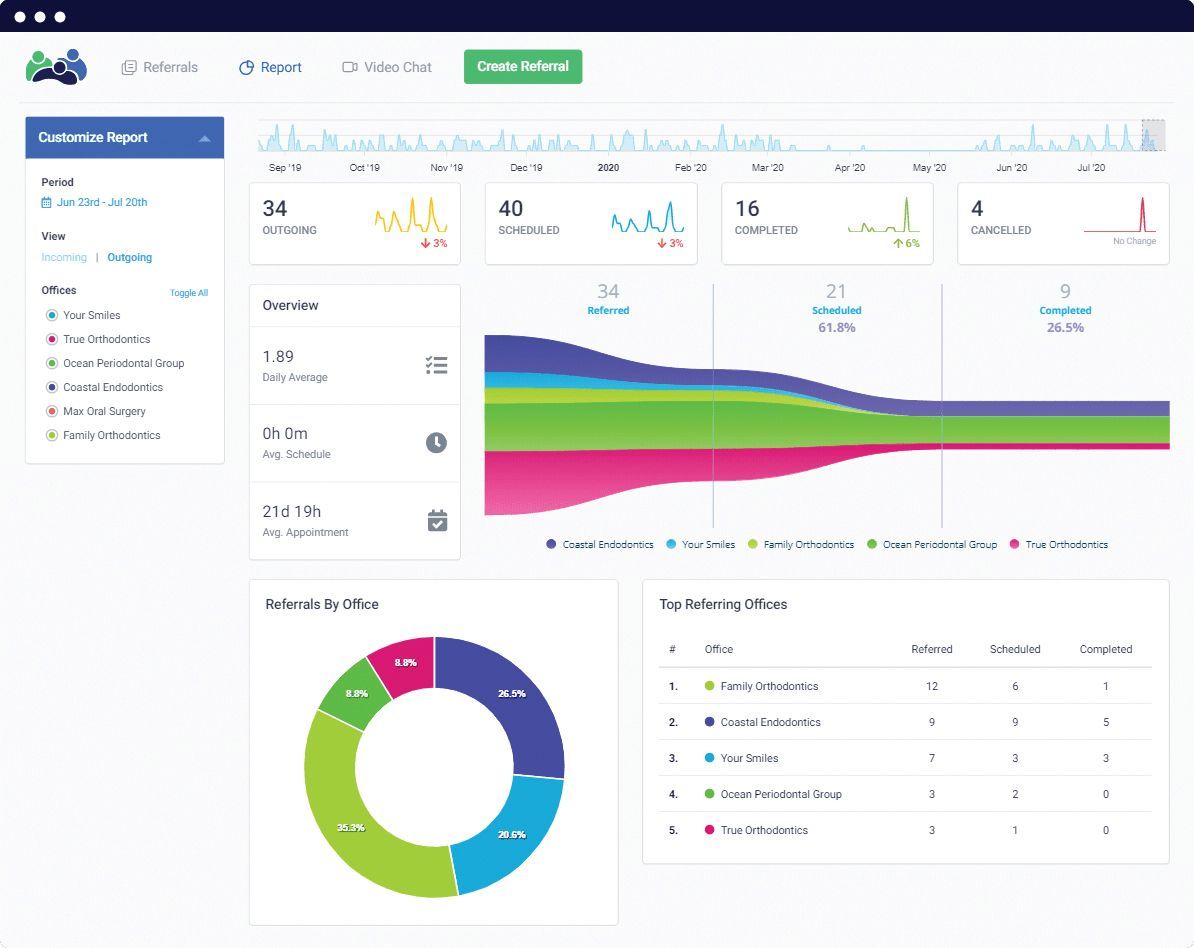
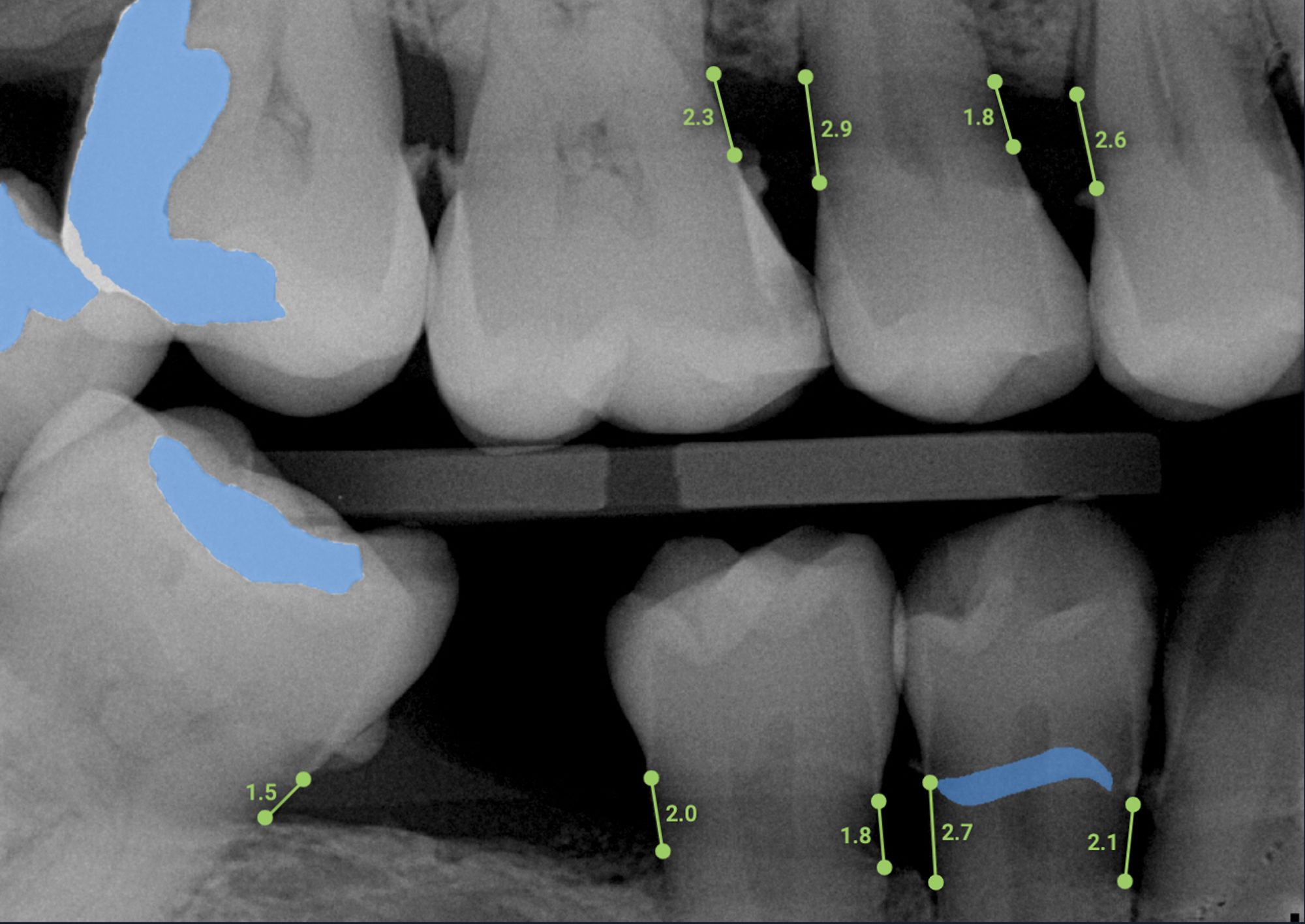
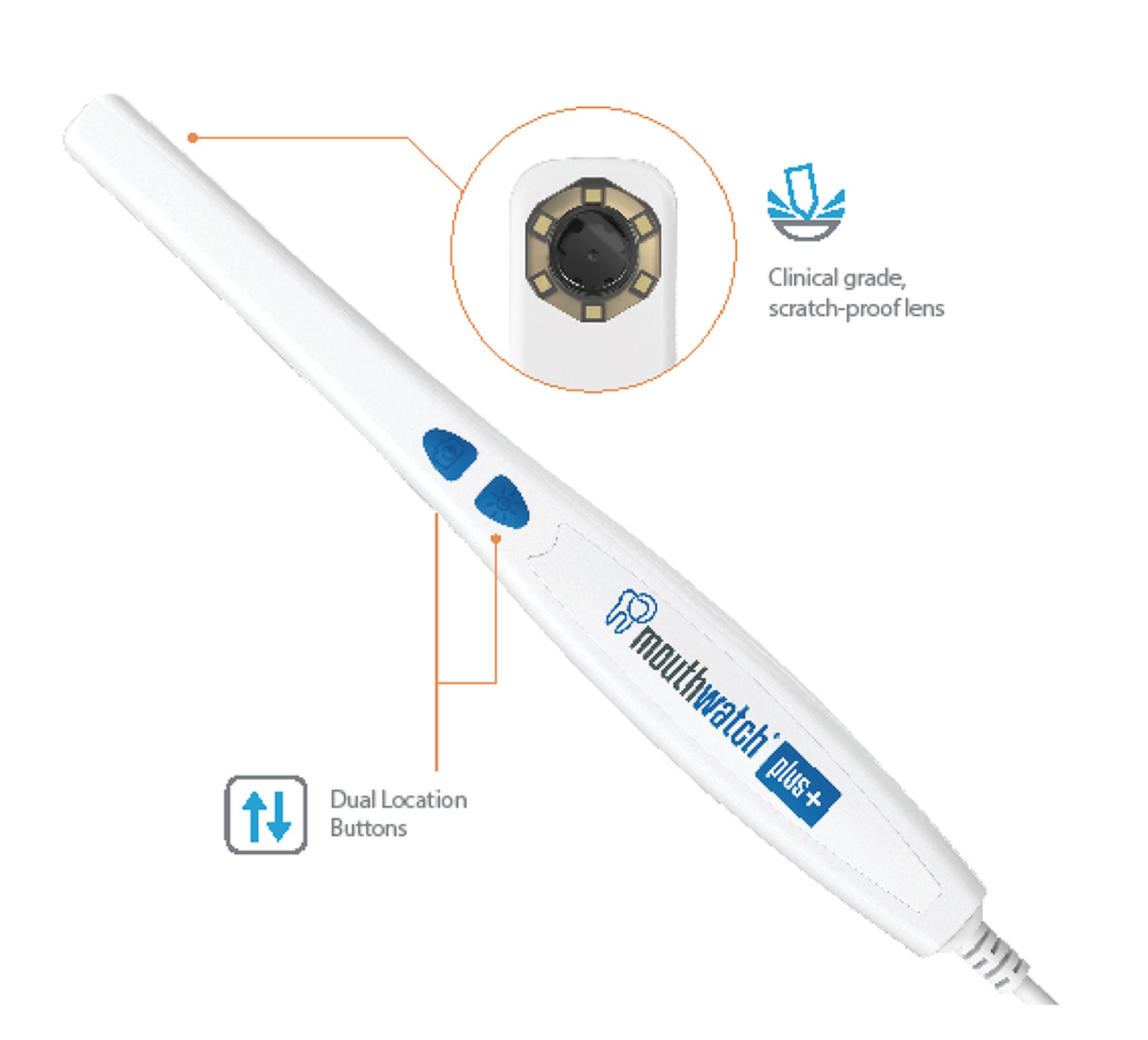
How often does a general dentist make a referral only to find out that the patient never bothered to make an appointment? How frustrated is a specialist’s front desk team member when they receive a random digital x-ray attachment from us only to be unable to find it in their inbox months later when the patient finally schedules an appointment? My favorite aspect of Refera is that every relevant attachment is tied to the referral form itself, meaning no more searching through old emails to find that x-ray. Another added benefit is the reassurance to my specialists that I am indeed supporting them more often than they may think—their office is notified when a referral is made (versus the old days of knowing I made a referral only if the patient scheduled with them).

As I’ve come to learn, the advantages of digital referrals are similar to those from making the switch to paperless everywhere else in my practice. For referrals, going digital allows me to track my outgoing treatments (which is very helpful for scheduling follow-up work), reduces my practice’s liability, increases staff efficiency, and ensures my patients receive the best care possible. I have formally banned paper referral forms at my office. When a representative for a specialist arrives at my office with a box of cupcakes and a stack of referral forms, I inform them that we use Refera exclusively with all our partner specialists because of how easy it is to use. Soon, those offices catch on to the benefits and often will thank me for the recommendation.

Refera is one of the best things to happen to my practice in many years, and my hope is that by sharing it here others can benefit as well. General dentists, specialists, and patients all win with a digital referral.


June 1, 2022

It’s Time to Embrace Teledentistry’s Hybrid Workflow

As a locum tenens dentist who works in practices that range from sole practitioner to group practices, urban dental service organizations (DSOs) to rural areas, I noticed that the day starts by reviewing the day’s schedule with the team.

TeleDent from Mouthatch

I introduced 2 of the offices I frequently work in to TeleDent, the all-in-1 teledentistry platform from MouthWatch. I also advised them to set up a separate teledentistry schedule for patients who could be served more conveniently with a virtual visit- which is a patient-selected option

This was achieved by booking 15-minute teledentistryappointments, either in-between in-office patients during operatory clean up or grouped together at the end of the day.

In this way, the dental team and I could maintain face-to-face contact with new and existing patients without PPE – which puts patients, especially children at ease! To participate in a virtual TeleDent consultation, all the patients need is a device such as a smartphone, tablet, or computer.

For new and existing patients alike, TeleDent can help in performing medical history and insurance information uptake. Virtual consultations can also include conducting limited exams. This way, when the patient does come into the office for a comprehensive exam, the chair time is optimized in the operatory.

As a follow-up to the comprehensive in-office exam, a virtual appointment can be immediatelyscheduled to review images, present the treatment plan, obtain informed consent. At the close of this virtual case presentation, anin-office appointment can be scheduled where chair time will befocused on performing the recommended treatment.

In both practices, the dental teams love the efficiency of TeleDent, and the patients love the convenience of not having to travel to the office for a conversation if they choose.

By implementing TeleDent, dental practices can now create a hybrid workflow consisting of virtual consultations and physical treatment. In my opinion, based on firsthand experience, this hybrid workflow model should be carefully considered by every dental practice - especially in the post-COVID era.

Since the introduction of new infection control protocols, it is no longer efficient or cost-effective to bounce from room to room. With TeleDent,more care can be provided for each patient, sometimes with longervirtual visits, often with pre-payment for services, which reduce the number of cancellations.

I predict that the “blended” teledentistry workflow will become the norm. When is hard to predict, but why wait to see what others do? Your patents and your practice can be benefitting from teledentistry’s “blended workflow” today. I’ve seen proof of this over the last 2 years.


June 1, 2022

Treat PRR, Small Class I Pediatric Cases With a Bioactive Restorative

Less is more when it comes to children’s oral health. This applies to pretty much everything one can conjure up, from snacking and sweetened beverage intake to the amount of toothpaste used in preventing dental caries. Minimally invasive dentistry is built upon the same rationale and seems to serve our youngest patients the best.

Keeping in mind the vulnerability and attention span of children, it is only fitting that we reflect on our choices of treatment modalities for the No. 1 dental disease that affects them early on in life.

Beautifil Kids SA

Beautifil Kids SA

Depending on the extent and severity of the caries and the patient’s development and ability to cooperate, treatment plan options can include silver diamine fluoride by itself or in conjunction with SMART restorations, conventional restorative treatments such as composite resins, glass ionomers, resin-modified glass ionomers, and stainless-steel crowns. The neat thing about providing dental care to children is that there is neither a 1-size-fits-all approach nor a single dull moment in our day.

As clinicians, we are constantly learning and exploring ways to make evidence-based dental treatment easier, faster, and more efficient for our young patients to ensure a lifetime of good oral health and positive dental experiences. And as practice owners, we are constantly mindful of incorporating materials and methods that enhance productivity and improve workflow while keeping overhead costs reasonably sound.

In the pursuit of this goal, a newer composite material checks all these boxes quite well; it fits in quite well in a fast-paced, multicolumn, multi-provider pediatric dental practice.

Shofu’s Beautifil® Kids SA is a comprehensive self-adhesive flowable restorative with bioactive Giomer Technology that eliminates the need for etching and bonding and can be used to restore teeth right after prep/caries removal. The elimination of these 2 technique-sensitive steps can revolutionize treatment for fidgety children. The primary indications are PRR and small Class I restorations. In conjunction with an isolation system such as Isolite or DryShield, it works wonders in the quality and the speed of the restorative treatment. Giomer Technology is a proprietary filler particle with healthful benefits that are incorporated into Shofu’s tooth-colored restorative materials.

Giomer Technology offers fluoride release and recharge, which is clinically proven to prevent caries recurrence. It also provides properties of composite resin such as excellent esthetics, easy polishability, biocompatibility, and smooth surface finish. Moreover, it forms a material film layer with saliva that is reported to minimize plaque adhesion and inhibit bacterial colonization


June 1, 2022

Choose an Open Architecture Workflow for Digital Dentistry

The decision to step into the world of digital dentistry was an easy one for me. Accuracy, efficiency, and patient comfort are just a few obvious benefits of a digital workflow. In addition to the advantages of digital scanning, a beautiful, exceptionally accurate restoration delivered within 15 minutes of prepping wins over a 2-week turnaround. But what many dentists don’t realize when selecting a scanner, mill or printer is that there is 1 choice that will impact how your practice functions and evolves: open versus closed architecture.

In an open architecture system, your scanned files are saved in a commonly used format and can be imported into any CAD software. This file can then be sent to any mill, printer or other hardware, which means you can send it to the lab of your choice or use it in-house for chairside production. In contrast, if your system has closed architecture, the digital patient records you create are compatible only with thatparticular scanner, software and mill included in the closed system.

Both systems deliver quality restorations, however, an openarchitecture system allows clinicians more restorative material choices, more lab choices, and most importantly the ability to adapt to the industry’s changes by adding new technology. For this reason, I chose Roland DGA’s open architecture DGSHAPE DWX-42W Chairside Milling Solution. The DWX-42W seamlessly integrated with my existing scanner so that I could easily and quickly jump into delivering quality, ultra-accurate chairside restorations to my patients. Its compatibility with other software also allows me to mill and deliver multiple restorations in one appointment - a massive benefit for my patients.

Choose an Open Architecture Workflow for Digital Dentistry

Choose an Open Architecture Workflow for Digital Dentistry

My current workflow consists of the 3Shape TRIOS Scanner, Trios Design & Implant Studio CAD Software, MillBox CAM Software, and the DGSHAPE DWX-42W Chairside Milling Solution. All the pieces of my workflow integrate seamlessly.

My advice to anyone who’s new to an open architecture workflow is to begin by producing 1 thing that’s simple. For most clinicians, that’s a single crown. My tips on this process are based on my own experience of doing the exact opposite. My first or second case was a 10-unit anterior esthetic case. I stumbled a bit, but it helped me learn and improve my process quickly. So, a few tips to consider:

  1. Start with a single crown – make it a posterior, not an anterior.
  2. Choose a patient with no temporomandibular Joint (TMJ) issues and no occlusal challenges.
  3. Give yourself more time initially so you don’t feel stressed or rushed – you will quickly become more efficient.
  4. Have your team communicate to your patient regarding the precision, accuracy, and speed of this system, so that the patient appreciates yourinvestment and elevates their perception of you as a clinician.
  5. Have fun - this part often gets missed but the simplified workflow and ability to integrate with other systems makes dentistry fun.

Remember,how your process looks to the patient is what makes a digital workflow your biggest asset and a game-changer for your practice. Simple protocols can make a huge difference when it comes to time saved, patient education, and best of all, the exceptional fit and esthetics of the final restorations.

My decision to choose Roland DGA’s DWX-42W open architecture platform came from one desire: control. I am always evolving as a clinician, a business owner, and a person, so I want to maintain my ability to choose the best technology options, now and in the future.


June 1, 2022

Use Artificial Intelligence to Enhance Diagnostic Precision and Practice Efficiency

With every passing day, more clinicians are using dental artificial intelligence (AI) to enhance their clinical decision-making, improve patient care and communication, and make practice performance more efficient.

During a recent visit to a general dentistry group practice that adopted Overjet’s AI technology, I had the opportunity to see how the dental AI product was being used by different team members to better serve their patients.

It was fascinating to see how the clinicians and team members— who ranged in age and roles—viewed dental AI, each through a different lens.

The experienced dentists? Two of the senior doctors who had been in practice for more than 30 years described the dental AI as an important clinical decision support tool. The technology also helped them standardize the quality and delivery of dental care in the practice as they brought new clinical associates into the practice.

The associate dentists? The newer doctors in the practice also saw the value of using AI to enhance patient communication and case acceptance. They recognized that Overjet is a powerful assisted technology that helps ensure all radiographic findings are efficiently, consistently, and accurately identified. Dental AI enables greater precision and accuracy in the entire diagnostic and treatment planning process.

Using AI as a diagnostic tool.

Using AI as a diagnostic tool.

The dental hygienist? By the time a patient steps into the office for an appointment, the dental hygienist has already preplanned much of what needs to be done. Using dental AI’s auditing feature, the hygienist has the opportunity to review the next day’s patients and identify what radiographs need to be taken and what information needs to be updated. AI enables the hygienist to have a much more efficient workflow. Visualizing caries, calculus, or bone loss on the x-rays helps the hygienist discuss disease processes and clinical needs with patients.

The care coordinator? A great advocate for dental AI in the practice was the care coordinator. Why? The care coordinator is the one who reviews the information and dental x-rays with the patient after the dental team has completed the examination, made the diagnosis, and offered treatment planning options. Dental AI enables the care coordinator to highlight the clinical problem areas and reiterate why the doctor has recommended certain treatments. This enhances patient understanding and case acceptance.

These team members’ experiences are great examples of how dental clinicians are using AI technology to improve care delivery. Modern practices are realizing the clinical and operational value of innovative technologies and moving toward smarter operations that leverage dental AI. As clinicians harness innovative approaches to ensuring the highest quality care in their practices, the applications for dental AI are only just beginning.


June 1, 2022

Erase Skepticism With Your Intraoral Camera

My intraoral camera is a great tool to help me educate patients. I wish that is all I wanted to use it for. But unfortunately, we need it to help correct some of the wrongs in our profession.

Recently I read about a dentist in Wisconsin who was convicted of cracking his patients’ teeth. According to an NBC news report, the dentist would show patients an x-ray of their tooth and point out a line or crack in the tooth, tell them it was a fracture or decay, and then use his drill to break a portion of the tooth, x-ray the tooth again, and submit the new x-ray to the insurance company for reimbursement.

Headlines about dental fraud or overtreatment charges linger in patients’ thoughts and make patients uneasy about what their dentists are doing. They don’t know. They cannot see what is going on.

But they can see—with our help and reassurance from intraoral photos. Many patients will believe what we tell them and agree to treatment easily. However, there are some who want to see exactly what is wrong with their mouth or want to know more about the procedure. I sense their skepticism and hear the hesitancy in their decision-making.

Instead of getting upset about the patient not believing me or taking it personally, I simply get my intraoral camera out to take pictures and show them what I am seeing. We can talk and explain, but nothing helps a patient understand and see their issues like a photo.

Figure 1

Figure 1

I use MouthWatch cameras to help me erase this kind of healthy skepticism. I took a picture of my patient’s cavity because it was located on the buccal of #15 (Figure 1). It was nearly impossible for the patient to see; I could not see it on the x-ray and I wanted to show my patient what was happening. He said he didn’t feel any symptoms and wondered why he needed the restoration. Then he saw the photo and realized the problem.

It’s very important to not take questions such as this personally. Some colleagues take offense when a patient questions their diagnosis. With today’s media options, patients see all kinds of messaging, and the bad messaging often gets the most clicks. The Wisconsin dentist story was all over the news. Patients remember these stories and may be skeptical at their next visit.

According to a 2016 Gallup poll on perceived ethical and honest behavior, dentists ranked fifth. With intraoral cameras, we can calm our patients’ fears, erase the skepticism they have, and continue to be a trusted profession for our patients. When patients trust us, they agree to the treatment we recommend. Case acceptance rises and patients receive the treatment they need.


June 1, 2022

Limit Contamination During Bonding with a Nonacidic Cleaner

Contamination can be a major contributing factor to restoration failure when bonding direct and indirect restorations. A surface contaminated with saliva and/or blood can jeopardize the bond strength, which can cause issues with adhesion, leading to debonding or complete restoration failure.

A common scenario is when removing a temporary crown prior to seating the permanent restoration. Temporary crowns tend to leak, resulting in contamination with saliva, blood, bacteria, and biofilm. Using a safe and effective cleaning agent is extremely critical prior to cementation, and my choice is KATANA™ Cleaner from Kuraray Noritake. It’s quick and easy to use. Apply and rub KATANA Cleaner on the surface of the restoration, abutment, or cavity prep for 10 seconds, then rinse, dry, and cement the restoration according to the manufacturer’s instructions.

Figure 1

Figure 1

Figure 2

Figure 2

With a pH of 4.5, which is safe to use both intra- and extraorally, KATANA Cleaner is an all-purpose universal cleaner for tooth structure and prosthetics (Figures 1-2). It works with virtually all restorative materials, including ceramics (zirconia, lithium disilicate, porcelain), resin-based materials (CAD/CAM crowns, composite resin), metals (precious and nonprecious alloys), and posts (glass fiber and metal), so there’s no need to stock multiple bottles of other cleaners when KATANA Cleaner will cover all your bases.

KATANA Cleaner is a nonacidic solution that will not harm or demineralize tooth structure. I think of it as a “dental soap” that’s simply cleaning the surface and nothing more—which is what you want in a cleaner. It is a nice substitute for a traditional pumice, which can get messy and cause gingival bleeding. I also prefer it over air abrasion, which may do a great job cleaning the tooth but may cause some bleeding that can negatively affect the stability of the bonding agent.


June 1, 2022

Obtaining Dry Field Isolation in Class V or Cervical Defect Case

A Class V or cervical defect can be unsightly and sensitive. It also can become deeper with time if left untreated. The defect is often a combination etiology: abfraction, recession, and abrasion. When restoring with resin material, clinicians should be precise in obtaining a smooth restoration that transitions well from root to resin and resin to enamel. Dry field isolation is absolutely crucial and can be obtained in several ways. I typically use the OptraGate (Ivoclar), the Ultrapak #000 retraction cord (Ultradent), and a soft tissue laser as needed if the margin is not accessible from retraction cord placement alone.

Obtaining Dry Field Isolation in Class V or Cervical Defect Case

Obtaining Dry Field Isolation in Class V or Cervical Defect Case

The material of choice for cervical defects is the G-aenial Universal Injectable (GC America) with an optional internal layer of color blocker (Tokuyama’s OMNICHROMA) for darker roots.

Step 1.Prepare dry field, lightly prepare enamel margin with a diamond bur. Optional: use a soft tissue laser for gingivectomy to access the cervical margin.

Step 2. Selective etch enamel surface for 15 seconds and rinse.

Step 3. Pack #000 cord in the sulcus.

Step 4. Prime and bond (G-Premio; GC America), air thin, and cure.

Step 5. Add color blocker (OMNICHROMA) layer in deepest aspect and cervical floor. Cure.

Step 6. G-aenial Universal Injectable of selected shade is then applied either directly from the syringe or indirectly with the tip of an explorer. This resin holds its form and can be moved around and shaped while keeping the explorer in the body of the resin before it is cured. With time, it becomes easy and there are no voids in the final restoration.

Step 7. Remove cord and shape and polish the resin as needed. Take care around the root to avoid creating sensitivity from overpolishing.

I find that moving the resin to shape it with the explorer gives me more control in small areas. It also has excellent adaptation to the root surface and high surface polish. It is easy to achieve a glasslike finish and smooth transition without having any ledges sticking out or voids between resin and cervical margin. This restoration tends to be smooth and therefore, kind to the tissues.


June 1, 2022

Reconsider Your Endodontic Referrals

In dental school, one of my biggest fears was that my endodontic therapy was not accurate. I wanted to ensure proper access and find all the canals without an unexpected perforation. It also was important to find the ideal length and instrumentation, confirm the proper length with the adequate gutta-percha point, and use the perfect sealer to have a beautiful final x-ray.

After almost 25 years of practicing as a general dentist, I still am anxious about that final x-ray, perhaps because, like many dentists, I always want to achieve perfection.

With time I learned that I prefer to treat relatively simple endo cases and refer to my phenomenal endodontist for all the complicated ones (sound familiar?). I also worked my way through different systems and techniques from hand instrumentation to rotary, traditional film x-rays that took an eternity to digital imaging, and a very accurate apex locator. However, I still prefer to see that measurement on an x-ray. Magnification also has improved what we can see, and now it is almost the standard of care. I even use my CT scan in some cases to confirm that I am satisfied with the final treatment. As a result of this journey, it’s easy to conclude that doing endo nowadays is a lot easier and even more predictable than in the past. But it is all for naught without a good sealer.

BIO-C Sealer Ion+

I have used a newer bioceramic endodontic sealer, Bio-C Sealer Ion+, and have been satisfied with the results and how it facilitates the entire process. Two bioceramic sealers (EndoSequence BC Sealer, Brasseler; Bio-C Sealer Ion+, Angelus) produced positive results in cytocompatibility, unlike resin-based AH Plus, and they demonstrated high calcium ion release and increased mineralized nodule formation in contact with periodontal ligament cells, according to results of a recent investigation.1

Advantages of Bio-C Sealer Ion+ include the ability to have the material ready with no need for mixing or manipulation, the direct insertion with a disposable intracanal syringe, and high flow. It has reduced my work time, and I no longer worry about not having reliable endo sealer material. And Bio-C Sealer Ion+ is approximately half the retail price of BC Sealer. As co-owner in 4 practices, that also counts.

Using Bio-C Sealer Ion+ gives me an easier workflow and facilitates the handling and predictability of my endodontic treatments to the point of rethinking my endodontist referral process.

References

  1. Sanz JL, López-García S, Lozano A, et al. Microstructural composition, ion release, and bioactive potential of new premixed calcium silicate-based endodontic sealers indicated for warm vertical compaction technique. Clin Oral Investig. 2021;25(3):1451-1462. doi:10.1007/s00784-020-03453-8


June 1, 2022

Essix Retainer Technique is Designed With Healing Abutment in Mind

In the majority of my dental implant surgeries, a healing abutment is inserted at the time of implant placement. My preferred method of removable provisionalization is a tooth-borne Essix retainer because, if properly designed and adjusted, it exerts no force on the healing abutment and implant during the integration period. I notice a lot of times that an Essix designed with an ovate pontic requires additional operative time for adjustment and proper seating. The adjusted result occasionally is less than ideal and the patient leaves with an unsatisfactory esthetic appliance.

Here is the unique workflow we use in our dental laboratory, Digital Provisionalization Technologies, for designing and fabricating a unique Essix retainer based on the anticipated healing abutment size and position determined from an X-Nav Dynamic 3D Navigation virtual treatment plan:

  • Step 1. The X-Nav virtual treatment plan (Figure 1) is imported into exocad DentalCAD software. The exported scan flag is detected and converted into a stock Ti-base and the anticipated final restoration is designed. The restoration and intraoral scan are saved as a single .stl file and printed on a NextDent 5100 3D printer with model material. The printed model is then light cured and finished per the manufacturer’s instructions (Figure 2).
  • Step 2. The designed restoration is divided—one part is the gingival third, which is saved and printed to be luted onto a stock Ti-base and used as a custom healing abutment (Figure 3). The other part is the incisal two-thirds, which is saved and printed to be used as the pontic in the Essix retainer. Both pieces are printed with NextDent C&B MFH material, light cured, and finished according to the manufacturer’s instructions.
  • Step 3. The retainer portion is created using the suck-down technique with 1-mm thick invisible retainer material. It is then trimmed and scalloped to the gingival margins with a hot scalpel blade.
  • Step 4. The printed pontic is brushed with a small amount of composite, inserted into the Essix, and light cured. A pinhole is placed with a fissure bur through the palatal side as an additional retention aid (Figure 4).

At surgery, the osteotomy is prepped and the implant placed using dynamic navigation assistance following the X-Nav virtual treatment plan. Once the implant is in proper position and any supplemental grafting has been performed (Figure 5), the custom healing abutment is inserted and tightened by hand (Figure 6). The Essix is fit and verified to be seated completely and not impinging on the custom healing abutment (Figure 7).

This technique has 2 main advantages over a conventionally made Essix retainer. First, very minimal, if any, operative time is required for insertion because it has been designed and fabricated to seat over the healing abutment. The second and most important advantage is the superior esthetic outcome that can be achieved due to the seamless appearance of the custom healer and Essix retainer. Offering this simple, cost-effective, esthetic, removable provisional option to our patients is a great way to differentiate our practice and optimize outcomes.


June 1, 2022

Prevent Postoperative Sensitivity With Antimicrobial Desensitizer

Postoperative sensitivity can be a problem for some patients despite all the advancements in bonding agents and techniques. You can consistently reduce your chances of creating sensitivity to a tooth that you have restored by using a desensitizer.

Hemaseal & Cide

Hemaseal & Cide Desensitizer from Advantage Dental Products is easy to use (it won’t annoy your dental assistant), doesn’t take a lot of time to apply (it doesn’t need to be light cured), performs more than 1 function at a time (it can multitask), and is not a financial strain on the bottom line (it’s one-third the price of other well-known desensitizers).

The superior antimicrobial activity of Hemaseal & Cide is due to the presence of 4% chlorhexidine. This not only disinfects the dentin surfaces but also increases the bonding strength by up to 33% over time. The desensitizer is hard on bacteria—it prevents microleakage around restorations—yet soft on the surrounding soft tissue, and it does not sting the gingiva upon contact. Just apply Hemaseal & Cide with a microbrush and remove the excess. By using the desensitizer, you greatly improve the chances of having a successful restoration. I use Hemaseal & Cide in any procedure that involves cutting into the dentin.

Whether I’m prepping teeth for crowns, veneers, onlays, or composites, the steps for using it are the same. The tooth is rinsed off and isolated. If I’m going to etch (selective or total), it’s done now. With the use of a microbrush and 1 to 2 drops of the desensitizer in a plastic well, Hemaseal & Cide is applied to the tooth. The dentin is scrubbed with the microbrush for 10 to 15 seconds, then wicked away with a dry microbrush. Leaving the Hemaseal & Cide slightly moist, the tooth then is ready for bonding or cementation. It is compatible with all bonding agents and cements.

The additional time taken to incorporate Hemaseal & Cide into your restorative routine is worth the positive long-term effects this product has on teeth and restorations. I want my bonding to improve over time and I want the teeth that I’m restoring to not be sensitive. Hemaseal & Cide helps me achieve those goals and more.


June 1, 2022

How to Use Thermoplastics for Provisional Crown, Bridge Fabrication

The fabrication of a provisional crown or bridge takes place before abutment preparation occurs. This is initially done, typically with impression material like polyvinyl siloxane or alginate. Polyvinyl siloxane is stable for a long period of time and is especially useful when a remake of the provisional is needed, however it can be costly. Alginate, while less expensive, cannot be kept stable for very long and is not useful when a remake is required. One alternative to impression materials is thermoplastics.

Matrix Buttons from Advantage Dental Products

The Matrix Button from Advantage Dental Products, Inc. is a thermoplastic disc that we find is an excellent low-cost option to traditional impression materials. We can use them individually when doing single crowns or by combining multiple buttons, multiple crowns, and provisional bridges can also be captured. It is simple and easy-to-use; we heat the Matrix Button in a hot water bath or by using a ceramic mug in the microwave. Once the matrix is sufficiently warmed, it turns from white to translucent. The translucency indicates its softened thermoplastic state and then I form it with gentle finger pressure around the teeth. I always capture at least one tooth on either side of the prepped tooth or teeth as well, to insure repeatable seating.

Once it has been formed around the teeth, the soft thermoplastic can be instantly hardened by spraying it with cold air, water, or both. It will be ready for removal from the teeth once cooling turns it opaque white.

Following tooth preparation, I like a bis-acryl composite such as Luxatemp, injected into the intaglio surface of the formed matrix and seated to place against the teeth. Typically, it’s set within two minutes, the Luxatemp provisional is easily removed from the formed Matrix Button, trimmed, polished, and cemented using a provisional cement. Should it prove difficult to remove due to undercuts, the matrix can be re-softened in hot water and peeled away from the provisional. In addition, because of its accuracy, very little if any occlusal adjustment is required saving time on this element of the procedure as well.

We can save this matrix until the final crown is seated; in case the provisional is lost or broken we can make another just that fast. I should also mention that a Matrix Button costs about $0.75, less than a typical mixing tip for my PVS impression material.

Overall, this is a material and technique that I would highly recommend to my colleagues.


June 1, 2022

A Simple, Economical Workflow for Fixed Prosthetics

This simple and economical technique has improved the profitability of my fixed prosthetics.

Matrix Temp

MatrixTemp® from Advantage Dental Products is a unique one-to-one dual cure composite for placement into a matrix to make a temporary crown. Unlike other products, it is not a ten-to-one catalyst /base mixture—this is a one-to-one mixture of catalyst and base. Previously these one-to-one mixtures could generate exothermic heat during the cure, but this does not happen with MatrixTemp. It also exhibits very little shrinkage on setting so that after forming in the mouth it can complete the final cure outside the mouth and still fit excellently.

When I switched from using powder-liquid acrylics many years ago I loved a lot of things about the Bis-acrylic composites. However, one thing I did not like was the thinner watery consistency, which I often found did not drive the crown margins as well as I would like. MatrixTemp is quite viscus it can be used in auto mixing tips, or it is so thick that I can mix it with a small spatula and place it in the matrix in this manner. No matter how one places it in the matrix, once there, the viscosity causes great adaptation everywhere; particularly in the margins that could be short with another material or less than ideal. The total set-time is about a minute and a half to 2 minutes. During that time a rubbery phase will occur when it is best to remove it and set on another surface for the final cure. My favorite shade is A2 which seems to lend itself to most applications; A3 and A1 are also options.

I prefer to use this material in a product called Matrix Buttons, also from Advantage Dental Products. These are a thermos-plastic matrix which, when softened and adapted to the tooth before the crown prep, makes a great easy matrix which I load with the MatrixTemp. Once hardened, we simply trim the margins on the gingival, seat the temporary and adjust occlusion. It has proven to be quite durable, and we rarely have temporaries fracturing.

Along with the advantages that appeal to me, another great advantage is cost. Compared to some of the other self-mixing temporary materials, MatrixTemp is about one-third of the cost. The Matrix Buttons are less than $0.75; amazing when I compared that to an impression tray and any vinyl polysiloxane. Plus, I’m in and out of the mouth in about 90 seconds.

I use these extensively for my single crowns and even three- or four-unit bridges in the posterior. Moreover, I use it also for anterior restorations particularly if I’m not looking for a bleaching shade. It comes out so smooth out of the matrix button impression that I rarely need to pumice it although that improves the appearance even more.


June 1, 2022

Going Digital Makes a Big Difference

It was March 2020, 2 days after the COVID-19 lockdown started, when a patient came in with a broken maxillary denture split down the middle.

3D Printing from Carbon 3D

Everyone was tense. We were wearing masks. And with the pandemic just underway, I had no idea what resources would be available. Was the lab open? How long would a replacement take to make? Would the patient be able to return? How long would the lockdown last?

I called my lab and fortunately, they answered the phone. They were open but running on a skeleton crew. I explained the broken denture situation, fully expecting to hear the patient would be without her denture for 5 to 7 days. Instead, the lab tech reminded me about my newly purchased iTero digital scanner and then instructed me to piece the denture back together, secure it with superglue, and take a wash impression. Then he said I could scan the denture and create a digital file that would be sent to the lab to fabricate a newly printed denture. Three days later, a new Carbon-printed denture was delivered to my patient and fit perfectly.

Since then, digital scanning and Carbon 3D printing have changed my attitude—and my patients’ attitudes—about dentures. Simply put, I no longer dread doing dentures.

Digital scanning and 3D printing have resulted in better outcomes for our patients. The fit of the denture is more accurate, which reflects fewer postdelivery adjustments, and the esthetics are beautiful and very natural. And because of the digital workflow efficiency, we can complete the job in fewer visits, which boosts our practice’s bottom line. This also benefits the patient by delivering a better product and greater satisfaction.

Traditional denture workflows require impressions and wax rims, and there always is an element of measurement uncertainty. These procedures frequently resulted in copy errors that would then equate to an end product that was less than optimal. During this process, multiple visits and multiple follow-up adjustment appointments were needed.

We have gone to digitally scan the existing denture, which creates a digital file capturing every detail. The lab then receives the file and uses a Carbon 3D printer to create a more accurate appliance that fits better than anything previously possible. Often these dentures can be completed in 2 or 3 appointments versus the past analog methods.

We have found that digital scans provide a precision level that means fewer sore spots and much less discomfort for patients. And with such a superior fit, these dentures don’t require denture adhesive.

Moreover, digital scans and 3D printing produce a better esthetic result—the teeth look natural. Analog denture teeth previously were often monoplane and dull. With digital scans and Carbon 3D printers, results are natural-looking cuspal occlusion with an accurate fit. Furthermore, the materials used in the printers add durability and strength to the dentures. This translates into better function and healthier outcomes for patients. In some cases, the difference can be life-changing.

Better outcomes also mean improved business productivity. By implementing a digital denture workflow, our improved bottom line is improved. The efficiency of using a scanner to create accurate data, in the end, reduces chair time.

To get results such as these, your dental practice must have a good lab partner with the right skills and 3D printing technology. This is definitely the wave of the future because it makes for happier patients—and happier dentists.


June 1, 2022

Robin the Robot Alleviates Pediatric Patients' Anxiety

As usual, it is a busy morning at the clinic. In between doing paperwork and seeing patients, I overhear…

“1…2…3…”

“Mom, where do I hide?”

“4…5…Ready or not, here I come!”

A dental clinic is not the typical place where hide-and-seek is played, yet I often hear this countdown at our office in Grapevine, Texas.

Robin the Robot from Expper Technologies

Children play with Robin the Robot while waiting for their dental exams.

You see, they are playing with Robin the Robot from Expper Technologies, our newest team member and the most charming and supportive robot you could ask for.

I first noticed Robin after stumbling upon a video where the robot was explaining dental procedures to a young patient. I instantly became curious, but what convinced us to bring Robin the Robot into our dental family were study results that showed after an interaction with Robin children’s stress was reduced by 34%, which resulted in a 40% decrease in the time of dental procedure preparation. Moreover, results of a recent study done by UCLA demonstrated that after an interaction with Robin 90% of families wanted to come back and see Robin again.

In addition, Robin helps us in other important areas such as attracting new customers and improving our social media presence, all due to increased patient satisfaction.

Robin prepares patients by explaining dental procedures with colorful animations and fun dialogue. This decreases children’s stress and anxiety and supports them to better cooperate with the clinic’s personnel.

On this particular morning, Robin was playing with a patient I will call Raphaela. Before Robin, Raphaela was always apprehensive when visiting us. It would take effort and time to make sure that she felt comfortable, but now she finds comfort in Robin.

I recall the first time that Raphaela met Robin. She was instantly captivated by the silly faces Robin made. She approached Robin and was greeted by a hello and an invitation to play a game. After playing with Robin, Raphaela slowly walked up to Robin and whispered:

Raphaela: “Robin, I’m really scared of the dentist.”

Robin: “Raphaela, there is nothing to be scared of! The dentists here are really nice!”

Raphaela: “Wait…do you go to the dentist here, too?”

Robin: “Yes! Dr Matthew is my dentist. He helped me when my tooth was really, really hurting. They took photos of my teeth and brushed them until they were super shiny, and then my tooth stopped hurting completely!”

Raphaela: “Was it scary when they took photos of your teeth?”

Robin: “Raphaela, can I tell you a secret? I was really, really scared because my tooth was hurting a lot, and I thought it would never stop hurting. But then Dr Matthew was able to fix it so quickly, it didn’t even hurt!”

Every young patient could use another friend at the dentist. Robin improves the perception of going to the dentist because it focuses on patient experience and care. And now Raphaela is always happy to come back to us and play with her friend Robin the Robot.

We’re excited about the new opportunities Robin has given us in customer service, patient care, and, ultimately, brand differentiation.


June 1, 2022

A Simple Device Can Offer Grinding Relief

We have all seen the signs—a patient presents with a multitude of symptoms; everything ranging from headaches, muscle fatigue, limited opening/tight musculature, abfractions along the gum line, worn teeth, as well as generalized tooth sensitivity. In many of these cases, bruxism tends to be the leading cause. And because most patients aren’t even aware they clench and grind their teeth, we are asked to diagnose and offer a solution to a problem they can’t understand, and it can be frustrating.

Grind Relief PRO

We dentists usually begin by recommending a plastic horseshoe “splint” for the patient that can be rather costly, include a lab cost for the practice, and require a follow-up appointment for the patient in a few weeks. We can send the patient away without even addressing their immediate discomfort in a timely fashion. Nor do we even know if the patient will even wear or, even worse, receive a clinical benefit from this new appliance. Moreover, with subsequent dental treatments, the patient may have an ill-fitting appliance that becomes unusable and requires a new one at yet another expense.

Now, dentists have an option to provide an appliance that can provide immediate gratification and relief without the need for impressions or even a return visit to the office. The GrindRelief PRO offers an affordable and convenient bruxism solution; an FDA-cleared thermoplastic mouthguard that can be fitted right in the office in just a matter of minutes, providing the patient with immediate therapeutic benefits. It treats and mitigates the effects of nighttime bruxism by preventing the occlusion of the posterior teeth, thus shutting down damaging muscle activity. For patient comfort and increased compliance, the mouthguard only extends to approximately the first pre-molar where it is lined with a soft, moldable thermoplastic layer inside a rigid plastic shell. We simply soften the thermoplastic material in hot water, fold, and place the tray in the mouth and ask the patient to bite to form to the teeth and remove it after the material hardens. The material can be reheated and re-fit as many times as needed to achieve the desired result. The GrindRelief PRO also provides the flexibility of being placed on either the upper or the lower arch.

In my office, we find ourselves using it to manage the TMJ/Bruxism patient, but we also use it a lot in our restorative cases; as an insurance policy for those patients who may have undergone extensive rehabilitation due to excessive wear and overall destruction. It provides us a sense of security in not only protecting what we have done but also continuing to establish a de-programming effect and keeping the damaging muscle activity at bay. If there are changes to the occlusion or a failed restoration that needs replacement, we can simply re-heat and re-form this device.

We have found this a truly comfortable, easily fitted and adjusted, economically advantageous appliance that is must-have in any dental office.


June 1, 2022

Thank you, Leonardo DaVinci

The ability to determine vertical dimension of occlusion has been a vexing issue for many of us practitioners. Anatomical landmarks such as the tip of the nose or prominence of the chin do not make stable points of reference. Both of these landmarks move and cannot be consistently charted over multiple appointments. In addition, knowing the vertical dimension of occlusion prior to determining it for fixed reconstruction or removable denture fabrication cannot be gained from these landmarks. This leads us to guesswork and/or trial and error, neither of which is reliable, but it’s the way most of us were taught.

Smart Craniometer VDOC

However, there is a repeatable landmark that can be used to solve this problem, using “hard point” relationships that have been published centuries ago. The vertical dimension of occlusion is also the same distance from the external auditory meatus to the lateral border of the outer orbit rim. Then, we simply subtract 3mm from this measurement to adjust for freeway space and you now have the correct distance of closed vertical dimension.

The method that I now use for recording this distance is a gauge known as the Smart Craniometer VDOC: Vertical Dimension of Occlusion Caliper, from Groman, Inc. The caliper initially takes a measurement from the external auditory meatus to the outer orbital rim. The VDOC has a built in 3mm offset that should fit snugly between the most anterior part of the undersurface of the mandible and the nasal spine with the teeth in occlusion. The (Patent Pending) Smart Craniometer™ improves on existing devices by eliminating the error-prone step of re-adjusting the probe for freeway space since the 3mm adjustment is built into the probe structure.

This measurement is repeatable and reliable and is useful in cases involving patients with cranial pain such as migraine headaches, sleep apnea, TMJ disorders and pain, and other symptoms related to a reduced vertical dimension of occlusion. We find this device easy to use and it is fully autoclavable.


June 2, 2022

Increase Cash Flow by Making It Easier for Patients to Pay

How your patients pay you may need a technique touch-up. One way to make it easier for patients to pay is through automated text-to-pay reminders. Imagine that you receive back the insurance explanation of benefits (EOB) and there’s a balance. Wouldn’t it be nice to be able to text the patient with a link to the invoice instead of having to send a paper bill? A study by Reviews.org found that 70% of Americans check their mobile phones within 5 minutes of receiving a notification. By embedding a payment link, your practice will get paid faster.

Simplifeye dashboard

If you’re still requiring patients to walk to the front desk to check out, consider adding a wireless card reader to enable chairside payment. Choose a payment processor such as Simplifeye that accepts Apple Pay, Google Pay, ACH, credit cards, and debit cards with tap-to-pay and chip-reader technology. Nobody likes waiting in a line. Make it easy to pay in the exam room or consult room as soon as they agree to treatment.

Another tip for increasing cash flow: give your patients the option to pay with cash or by credit card with the credit card processing fee clearly displayed. Just as gas stations show both prices, health care providers should too. By having patients pay the credit card processing fee on their transactions, practices can save thousands of dollars every month.

You also can increase revenue by increasing case acceptance rates. One of the biggest reasons patients don’t move forward with treatment is due to cost. Imagine running a soft credit check, receiving an instant thumbs up or thumbs down notification, and being able to offer a monthly payment plan with confidence.


June 2, 2022

SmartMirror is 21st Century Technology for 21st Century Challenges

SmartMirror

The first patent for the modern mouth mirror was filed in 1894. At the time, the fastest communication was the telegraph.

Over the course of my 20+ year career as an orthodontist, there have been a few technological advances that have truly impacted my practice. They mostly addressed issues like imaging and clinical efficiency.

SmartMirror is tackling perhaps the most overlooked issue in our practices— how to transform the century-old intraoral mirror design into a high-tech examination and patient communication device. SmartMirror incorporates world-class microimaging with powerful LED lighting to create a game-changing doctor and patient experience.

Video captured by the HD imager is streamed, in real-time, onto a chair-side screen for easy viewing. I like to see the video in my new patient exam room on a large ceiling-mounted screen, so I can include the patient and family in the process. This lets me be a doctor showing the benefits of treatment, not a salesman. Images and videos can be easily shared with referring doctors, saved to the patient file, or airdropped to the patient’s phone. The built-in microphone allows for audio recording. Intuitive zoom allows you to get up to 3mm from a tooth surface for up to 10x magnification. All of this while using a mirror with a world-class reflective surface that’s virtually scratch-proof.

Back pain? Neck strain? SmartMirror allows you to spend more time sitting upright, examining treatments on the chair-side screen instead of hunched over like a pretzel.

All of the above are amazing features. But what are the benefits?

Relationships

Our success as clinicians and small business owners depends on quickly building trust with our patients and letting them accept our recommendations for the treatment that they both need and deserve.

Whether it’s a new patient exam, or a hygiene recall, by letting the patient be part of the process, by letting them see, in real-time, what we see, we can build trust. We don’t need to sell dentistry. We simply need to let the patient see with their own eyes.

SmartMirror is not an intraoral camera. SmartMirror is the instrument you use at every patient exam. It’s comfortable to use and has a great ergonomic feel. You use it like any other mirror, but when you want to examine a prep, document treatment by recording a video or educate your patient about oral hygiene, it’s ready. SmartMirror puts 21st-century technology in your hand all day, every day.


June 2, 2022

Telehealth Takes Your Practice to the Next Level

As a young Egyptian immigrant, I struggled with a lack of access to quality medical and dental care. As a result, I’ve made it my mission to help bridge the accessibility gap in the dental industry. As a trusted dentist in my community, I often utilize telehealth services to provide the best care for all of my patients, regardless of their personal circumstances. Using modern innovations in telehealth to manage care reduces costs and provides a safe, convenient experience for patients who may not have had access to these services. There are often cost barriers, time constraints, and limited access to an orthodontic or dental office. Providing affordable, convenient care and alternative options empowers patients to make the best decisions for their unique needs.

Although teledentistry has been around since the 1990s, it wasn’t widely adopted until the COVID-19 pandemic, and research supports its increasingly important role in delivering safe, effective oral care in the future. According to a recent consumer survey, of those who had a telehealth visit in the past 2 years, 77% had a neutral to positive experience, and 59% agreed or strongly agreed that they were more open to receiving care through telehealth as a result of the pandemic.1

What’s more, 60% of survey respondents believed an affordability gap exists in the health care and dental fields.1 Too many individuals in underserved populations go without ever seeing a dentist in their lifetime, and teeth straightening treatments are out of reach for many patients. That’s why my practice joined SmileDirectClub’s Partner Network—to increase access to quality dental care and promote more diversity in the dental industry. Since joining the Partner Network, I have been able to boost patient access, grow my customer base, and expand my existing clear aligner services. The streamlined process of the Partner Network also made it easy to integrate into my practice without any overhead.

SmileDirectClub’s hybrid aligner model allows dentists to provide access to orthodontic treatment to a wider range of patients, including those who may not have been able to afford traditional orthodontic treatment, who have had relapses from prior orthodontic treatment, or who simply do not have the time for frequent visits to the dental office. After a patient’s initial office appointment, information is submitted to SmileDirectClub’s telehealth platform where their affiliated network of state-licensed doctors assesses the patient’s candidacy for clear aligners and, if appropriate, prescribes and monitors the treatment from beginning to end. Patients can review their treatment plans, and all of the aligners are directly mailed to them in 1 shipment. There’s also a mobile application that monitors and encourages adherence, and regular telehealth check-ins are scheduled to ensure that the treatment progresses safely and as planned.

SmileDirectClub’s Partner Network gives my practice the ability to offer a best-in-class option to those who have mild to moderate misalignments without sacrificing profitable chair time or in-office production. Patients receive a more convenient, affordable, and accessible option to achieve their desired smile. By offering telehealth services, dental providers can bridge the access-to-care gap with innovative solutions that break down barriers for patients to receive dental care. Elevate your practice by embracing telehealth.

Reference

  1. SmileDirectClub survey reveals growing popularity and acceptance of telehealth. SmileDirectClub. March 24, 2022. https://investors.smiledirectclub.com/news-releases/news-release-details/smiledirectclub-survey-reveals-growing-popularity-and-acceptance


June 2, 2022

This Should Be in Everyone's Toolbox

After 40 years of practicing dentistry, utilizing general anesthesia and sedation, I often look back at that snapshot of dental school and think about all I’ve learned afterward. So many clinical and business tricks, tips, and time-saving solutions to pass along before I retire.

I’m more of a form and function kind of clinician; envious of the classmate or colleague who can place and sculpt that ideal posterior restoration. A good friend of mine and McGyver-type dentist showed me how to use the Occlusinator® PRO Composite Finishing System. Even in my hands, the system allows me to create beautiful anatomic carvings in seconds and restore a tooth to ideal morphology and occlusion.

This simple system utilizes a Posterior Packer Sculptor (PPS) instrument for the bulk shaping. On one end of the PPS are two rounded packers measuring 0.8mm and 1.5 mm in diameter. The other end has a robust ball burnisher opposite an “acorn” burnisher, modeled after the famous 21B Acorn burnisher we used in dental school. That burnisher is based on the planar angles of the cusps on posterior teeth. Pack, burnish, and let the acorn form the planes with one instrument—no passing instruments, simply flick the wrist.

Once the composite material is cured, “connect the dots” formed by the acorn burnisher on the occlusal surface with a drag-and-drop motion. All 5 burs have a Safe Edge to control depth and prevent over-carving. The set features 3 acorn-shaped burs in large, medium, and small sizes that match the angulation of the acorn burnisher on the PPS for easy, repeatable composite sculpting. The acorn burs form the planes that comprise the triangular ridges and even the oblique ridges. There are two X-Mas Tree-shaped polishing burs that are coated with 15 – micron diamond particles that streamline the final finish of the planes and grooves. I rarely need any further polishing. Imagine that!

In a nutshell, the Acorns are for finishing, the X-Mas Trees are for polishing.

I estimate this system reduces my composite sculpting time by 50%. The burs’ self-limiting and Safe Edge gives me maximum control without skipping or scarring the enamel.

Every job or repair can be done better, faster, and easier with the right tool. The Occlusinator PRO Composite Finishing System should be in everyone’s toolbox.

Here’s how:

  1. Allow the appropriate-sized acorn to cut a “pilot” pit in the deepest landmarks.
  2. Like a child’s dot-to-dot drawing, simply go ½ way to next dot, then return ½ way from that dot.
  3. Avoiding cutting a straight path from mesial pit to distal pit preserves the triangular and transverse (maxillary) ridges so critical to supportive occlusion.
  4. This is identical to using a 21 B acorn carver when we do amalgams or soft sculpting composite.
  5. Quick, easy, accurate! Slight overfill will guarantee good occlusion.


June 3, 2022

Eliminate Cost as a Barrier to Treatment

Dental Products Report's Technique Touch-Ups 2022

For decades, payment assistance options for patients have been limited to a handful of prime-focused finance companies that only approved patients with high credit scores. For some of our patients—especially those who have difficulty paying for necessary treatments such as fillings, dentures, or crowns not approved for these options—it was becoming more difficult for them to afford the care they desperately needed. As dental providers in our community, we had to find creative ways to help, so we started a nonprofit organization that offers free dental care to help patients who couldn’t otherwise afford it. But many of our patients still went untreated. Dental offices across the nation are constantly looking for new ways to help their patients get the treatment they need without breaking their budgets.

Understanding Your Patient’s Payment Needs

From the moment a patient walks into our office, we’re doing everything we can to provide high-level service and care. That includes offering a variety of payment options. For too long, when patients were presented with treatment plans cost was often a barrier, with many choosing to walk out of the office without getting dental care they needed. We work with our patients to ensure they’re able to complete necessary treatments by helping them navigate their insurance coverage or finding them other payment assistance options. We want every patient under our care to leave feeling better than when they came in, and that includes fitting dental treatment payments into their monthly budget.

Find Financing That Serves Most Patients,
Not Just the Privileged

Dental patient financing has not changed over the past 20 or 30 years, and today less than half of applicants are approved through traditional options. That leaves many patients on their own trying to afford the care they need. In our experience, traditional financing plans are cumbersome with long application forms and processes. They often include lengthy approval and payment periods, which can delay treatments and test anyone’s patience. So we needed multiple financing options, including various prime and subprime offerings to serve our wide range of patients. Still, even with multiple options many patients walked away with embarrassing declines or were too overwhelmed with the application processes to start treatment. We had to have a better option.

Consider Alternatives Such as Buy Now, Pay Later

In 2020, we selected Sunbit as our preferred Buy Now, Pay-Over-Time provider, and it has been a game-changer for our patients as well as our staff. With Sunbit, we’re able to help 90% of our patients find an affordable payment plan with reasonable interest rates and higher maximum amounts than what we were offering previously. And we can do it all in approximately 30 seconds—no more long lead times on approvals or payments. With 9 out of 10 of our patients being approved by Sunbit and subprime and prime financing all in 1 platform without the hassle of multiple applications, what else can you ask for?

Our staff is confident in offering Sunbit’s service because of the high approvals and its easy-to-use digital application. The best part is that we’re paid in full the next day and we are not charged if our patients default. We’re seeing some of the highest patient retention rates in years. We’re able to offer more premium dental treatments, including dental implants as well as other important and necessary procedures, which is improving the overall quality of life for our patients. It’s a win-win for everyone.


June 3, 2022

Take Advantage of a New 3D Imaging System For Intraoral Imaging

Dental Products Report's Technique Touch-Ups 2022

Intraoral radiographs, bitewings and periapicals form the backbone of radiographic procedures in most dental practices. While the technology has changed with the advent of more efficient x-ray sources and digital detectors, the imaging geometry is basically the same as the first dental radiograph taken in 1896.

Basically, we are collapsing a 3D object into a 2D plane which degrades our ability to detect disease and abnormalities of the teeth and supporting bone. Closed contacts, superimposition of hard tissues and “cone cuts” are examples of the limitations of 2D imaging as practiced in dentistry today. Cone Beam CT (CBCT) has provided 3D information proven to increase our ability to detect disease and abnormalities as well as to treatment plan various maxillofacial and dental procedures.

However, metal artifacts lesson the usefulness of CBCT for dentoalveolar imaging. The higher costs and radiation dose of CBCT is also a concern of many dental practitioners. Some have wondered if we are at the end of the road for 2D dental alveolar radiography in terms of improvements in diagnosis and treatment planning. Is this true and is there an available intraoral 3D system that has the appearance and resolution of 2D radiographs?

The answer to both questions is yes. There is a new 3D intraoral system with the quality and resolution of 2D radiography without most of the metal artifacts seen in CBCT. It is called stationary intraoral tomosynthesis or sIOT.

I know that is a mouthful but let me explain the concept. Humans have 3D vision because our eyes are set apart. We are born with 2 light detectors from different angles. Imagine an x-ray system with 7 carbon nanotube sources and a digital detector which produces 7 radiographs taken from different angles almost simultaneously at a combined dose comparable to what is produced by today’s radiographic techniques. Now imagine that those 7 radiographs can be reconstructed in an image stack parallel to the detector allowing a dentist to “slice” through the tooth 1 layer at a time better visualizing the dentition and surrounding bone.1 Such image stacks would virtually eliminate closed contacts and better visualize cracks in the tooth or even root fractures that are not visible in intraoral radiographs or CBCT scans. Caries and periapical lucency detection could be improved as well as and the assessment of bone around implants and canal third/molar relationships.

That “imagined” system is sIOT and it is here today in the Portraytm imaging system by Surround Medical Systems out of Morrisville, NC. The technology was produced at UNC-Chapel Hill in a collaboration between the Department of Physics and the Adams School of Dentistry and is currently the only system using sIOT technology on the market. A few example images can be seen in the photo above.

References
1. Stationary intraoral digital tomosynthesis using a carbon nanotube X-ray source array. Shan J, Tucker AW, Gaalaas LR, Wu G, Platin E, Mol A, Lu J, Zhou O. Dentomaxillofac Radiol. 2015;44(9):20150098. doi: 10.1259/dmfr.20150098. Epub 2015 Jun 19. PMID: 26090933
2. Stationary intraoral tomosynthesis for dental imaging. Inscoe CR, Wu G, Soulioti DE, Platin E, Mol A, Gaalaas L, Anderson R, Tucker A, Boyce S, Shan J, Gonzales B, Lu J, Zhou O. Proc. SPIE 10132, Medical Imaging 2017: Physics of Medical Imaging, 1013203 (March 9, 2017); doi:10.1117/12.2254632.
3. The role of stationary intraoral tomosynthesis in reducing proximal overlap in bitewing radiography. Mauriello SM, Broome AM, Platin E, Mol A, Inscoe C, Lu J, et al. Dentomaxillofac Radiol 2020; 49: 20190504. PMID: 32202939 PMCID: PMC7719863 (available on 2021-12-01) DOI: 10.1259/dmfr.20190504


June 3, 2022

A Slam Dunk for Class I, V Restorations

Dental Products Report's Technique Touch-Ups 2022, Tokuyamas OMNICHROMA

I have a family practice just outside of Boston, Massachusetts, and here’s a technique tip that I use every day that makes doing some restorative procedures efficient, productive, and enjoyable: Class I, V, and incisal edges are a slam-dunk combo.

It’s an easy workflow because I only need to carry in stock the OMNICHROMA Flow flowable composite and the OMNICHROMA Blocker Flow (Tokuyama Dental America), which means I do not have to use my shade guide to try different shades. I used to pick a shade and then cure it onto the tooth to test and see if it was a good match and then use it or flick it off and try another. This was time-consuming and I had to keep an extensive inventory of shades. Sometimes they were expired or you couldn’t find the shade you needed.

But with fillings that are not deep—such as Class I, V, abfractions, and incisal edges—I go into it with a game plan. I will tell the patient that we can do this with no Novocaine, no shots, and no drill. There may be some smoothing and polishing at the end, but the air abrasion is like air and sand, and sandblasts the decay and creates retention. I’ll blow some air with my air-water syringe on the tooth and say it’s going to feel like this but sandy. Boom—happy and relaxed patients.

Now, this is only for some cases and some patients. Other patients may want you to get them numb regardless because they have super-sensitive teeth or are nervous.

Using air abrasion and OMNICHROMA Flow is also a great option for kids with occlusal decay. This won’t work for interproximal.

So, my protocol for a Class V abfraction with dark root or incisal edge or Class I caries, for example, is to insert the isolation device to retract, suction, and illuminate the area. Then air abrades the area. Again, no local or drill is needed. If the gum tissue starts to bleed, especially for gum line abfraction areas, then I’ll use some hemostatic agent or some cord to control moisture.

I will use bond and then cure and cover all dark or dentin with the blocker and cure and then regular OMNICHROMA Flow and cure. I use a high-intensity light so it takes just a quick few cures. I make sure when I add the flowable that it is in small controllable increments so it doesn’t run all over the place. Although the OMNICHROMA Blocker Flow and OMNICHROMA Flow are easy to handle, the less material and more control you have with the initial increments lead to less cleanup at the end.

I used to use the OMNICHROMA packable material but the flowable and the flowable blocker have become my go-to materials because of their esthetics and easy handling. If there is an incisal edge involved then I will typically use the packable version of OMNICHROMA Flow and blocker along with etch and a bevel.

You will have happy patients who say, “Wow, that was a lot easier and faster than I thought.” And you will be happy because you had a game plan. Do this over and over and you will get predictable results in a quick amount of time.


June 3, 2022

Connect With Dental Manufacturers on Your Time

Dental Products Report's Technique Touch-Ups 2022

Clinicians always wish they had more time. Between treating patients, completing administrative tasks, and striving for a balanced life, we all would like more time. vTail is a free app that connects clinicians with dental manufacturers on our time.

There are several companies on the platform (and more joining each month) who are able to answer our questions via a HIPAA-compliant phone call, text, or a video chat. Companies on the app will not reach out to the clinician, only the clinician will be able to connect with the companies of their choice. So, no unsolicited requests for information.

After linking to a company, the app automatically displays your area representative (even if you have never used that company before). One of the manufacturers on the platform is Kettenbach. Once we signed up for vTail, and connected to Kettenbach, our area rep Tom was listed. I am lucky. I have known Tom for decades and he is always available when I have an impression question. He is one of the rare reps who has my cell number. Many clinicians, especially women, prefer not to give out their cell phone numbers.

With vTail, everyone has the “Tom” experience. When we need something, or have a question, we are able to connect with one of the reps on the vTail app to answer our questions: all on our own time and without anyone having our personal contact number. This is not a selling app—no one will solicit us to buy products, but it definitely makes it easy to order if you need something last minute, without fumbling for a customer service number or your rep’s card.

The benefit of this app: the time you are able to save by avoiding going through a customer service telephone “tree” to get to the right person or technician to answer your question. The contacts are all in your hand. The app includes product information, IFUs, product specifications, and instructional videos.

The free vTail app has provided us with quick connections, solutions, options and informatics. I may now devote better time to my team, my patients, and my non-clinical life because my distractions with respect to product offerings and orders are lessened. I’ve also learned quite a bit about new dental manufacturers and service providers that I may not have had the opportunity to explore…and I can do it on my time table. This is perfect for a woman clinician who wants to spend time outside of practice life but still be in control of her practice.

In our practice, we refer to the vTail app as the "easy button”. Easy connection to the products and the reps we need, giving us time for quality dental care and a more relaxed quality of life outside the office. We are lucky. Everyone doesn’t have a Tom on speed dial. This places all your reps on speed dial without you doing a thing. vTail is truly something we didn’t know we needed until we need it.


Related Videos
CDS 2024: Diving Deep on J. Morita's New Root ZX3 & HF Module
CDS 2024: What's New at TAG University? with Andrew De la Rosa, DMD
CDS 2024: Breaking Down Barriers to Care with Eric Kukucka, DD
Contemporary Cosmetic Dentistry – Part I: Closing Black Triangles
Greater New York Dental Meeting interview with Robert Rosenfeld, DDS from Tokuyama Dental America
Greater New York Dental Meeting 2023 – Interview with Len Tau, DMD
Related Content
© 2024 MJH Life Sciences

All rights reserved.