Technique: How to cut procedure time in half with Tetric EvoCeram Bulk Fill
Placing composite restorations in the posterior region can be a time consuming and technique sensitive procedure.
Placing composite restorations in the posterior region can be a time consuming and technique sensitive procedure.
Historically, clinicians have used traditional direct composites to complete these restorations, carefully layering, sculpting and curing 2 mm increments. This process, while still used, presents several difficulties to the clinician, primarily in the form of polymerization shrinkage and shrinkage stress.1
These difficulties can cause stress fractures at the margins, marginal leakage, or an increased likelihood of secondary caries and post-procedural sensitivity,2 ultimately leading to clinical failure of the restoration.
The continued search for a more efficient and less technique sensitive procedure led to the invention of bulk fill composites. These materials allowed clinicians to fill and cure preparations up to 4 mm deep in one increment. Unfortunately, these materials were highly translucent and were available only in limited shades that appeared grey next to natural dentition.
Some of these materials also had a high reactivity to ambient operatory light; this severely limited the amount of time the clinician had to perform the restorative procedure, and the esthetics were compromised.
New Bulk Fill Materials
The next logical progression was to create a bulk fill composite that still exhibited high strength characteristics but had greater esthetics and manipulability.
This was achieved by altering the size of the filler particles. Modern bulk fills are typically nano-hybrids, meaning the size of the filler particles are much smaller.
This allows for a bulk fill material with a greater depth and uniformity of cure, a smooth consistency, life-like optical properties, and easier sculpting of anatomic form without requiring an additional “capping layer.”3
Tetric EvoCeram Bulk Fill is billed as a true advancement in bulk fill technology. One of the innovations featured in this product is the inclusion of the patented light-initiator Ivocerin™.
This new germanium-based light initiator allows a complete, homogenous cure in 10 seconds using any basic curing lights. One of the components of Ivocerin is an ambient light immunizer that ensures ample working time for the clinician to create artistic anatomical form.
Tetric EvoCeram Bulk Fill also contains a shrinkage stress reliever, eliminating the worries of shrinkage stress, because forces are evenly distributed across cavity walls and surfaces. Volumetric shrinkage of the material is very low, which provides a more predictably placed restoration.
The layered silicates that form the bulk of this material provide a smooth consistency, which facilitates optimal adaptation to cavity walls and easy contouring with conventional dental instruments. The following presentation demonstrates two effective bulk fill methods for posterior composite restorations compared with traditional layering and the time required to complete each procedure.
Case presentation
A healthy 45-year-old male patient presented with occlusal amalgam fillings that were placed more than 20 years previously. A treatment plan was agreed upon involving the restoration of tooth Nos. 30 and 32 using a Tetric EvoCeram® Bulk Fill composite, and tooth No. 31 using both a flowable and universal composite (Tetric EvoFlow® and Tetric EvoCeram®).

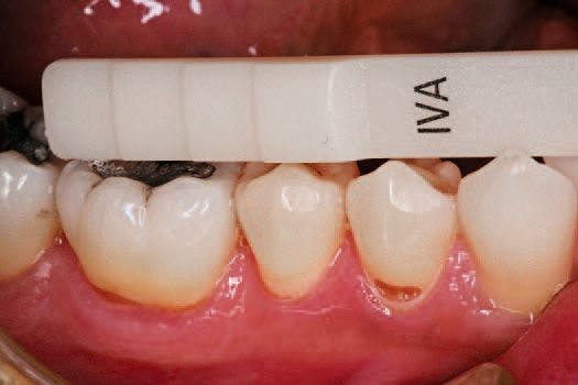

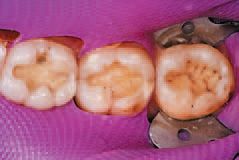
One of the three universal shades most closely matching the patient’s dentition was selected (Fig. 1). This patient was well matched to shade IVA (for slightly reddish teeth).
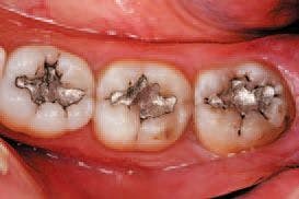
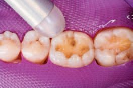
After inspecting the old amalgam restorations (Fig. 2), a rubber dam was then placed (Fig. 3). Next, using Sybron Dental’s Axis course diamond bur, KS3, the amalgam restorations and all damaged tooth structure were removed (Fig. 4).


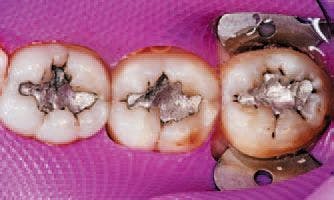


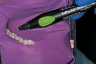
All remaining sharp edges of the preparation were then beveled with Sybron Dental’s Axis fine finishing diamond #846-016, completing the preparations (Fig. 5). Ivoclar Vivadent’s AdheSE® One F self-etch dental adhesive was applied to tooth No. 30 using the VivaPen™ delivery system (Fig. 6), leaving a shiny bonded surface (Fig. 7).


A stream of oil-free, moisture-free air expelled from a warm air tooth dryer from A-dec was then directed over the adhesives to evaporate the solvent (Fig. 8). The adhesive was light cured with Ivoclar Vivadent’s LED curing light, Bluephase Style, for 10 seconds.

Continued on the next page ...
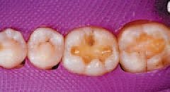
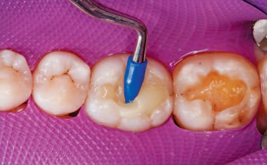
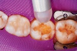
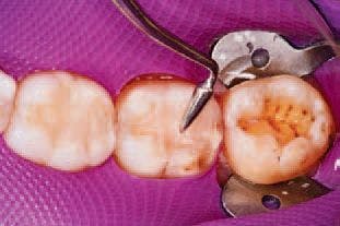
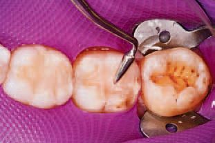
A single increment of Tetric EvoCeram Bulk Fill in shade IVA was then placed in the preparation of tooth No. 30 (Fig. 9), and shaped first with an OptraSculpt® sphere attachment (Fig. 10), then with an OptraSculpt pyramid attachment (Fig. 11). The final anatomy was contoured using a P1 plugger (Fig. 12). As a final step before finishing, the restoration was light cured with the LED curing light (Fig. 13).


Preparation then began on tooth No. 31. Ivoclar Vivadent’s Total Etch was applied to the enamel and allowed to penetrate for 15 seconds, after which the etchant was applied to the dentin and allowed to penetrate for 10 seconds to ensure proper etching of both surfaces (Figs. 14 and 15). The etchant was then rinsed off with water, and all excess moisture was removed.


ExciTE® F total-etch dental adhesive was then applied to the preparation and agitated for 10 seconds (Fig. 16). A stream of warm air was expressed onto the adhesive to evaporate the solvent (Fig. 17), and the adhesive was light cured using the Bluephase Style for 10 seconds.


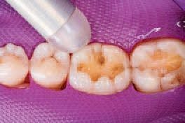
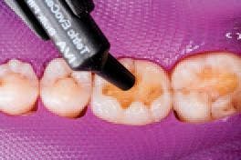
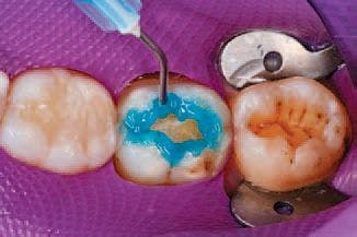
Tetric EvoFlow shade A3 was flowed 0.5 mm thick onto the floor of the preparation of tooth No. 31 and light cured for 10 seconds (Fig. 18). Then, a layer of Tetric EvoCeram shade A3 was placed within 0.5-1 mm of the cavosurface margin.


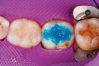
This second layer was shaped to replicate the dentin layer, then light cured for 10 seconds (Figs. 19 and 20).


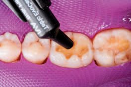
Tetric EvoCeram Transparent was then used to create each of the four triangular ridges of the natural tooth, and each ridge was subsequently light cured for 10 seconds (Figs. 21 and 22).


Lastly, preparations began on tooth No. 32 by applying the Total Etch phosphoric acid to the enamel and allowing 15 seconds for penetration, then applying the etchant to the dentin and allowing 10 seconds for penetration. Then, the etchant was completely rinsed off, leaving the area moist. ExciTE F dental adhesive was then applied, and a warm stream of air was directed over the preparation to evaporate the solvent.
Continued on the next page ...
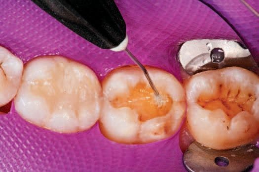
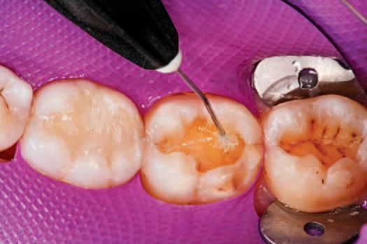
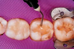
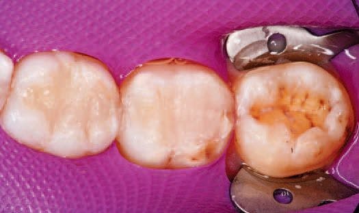
The ExciTE F adhesive was then light cured for 10 seconds. One increment of Tetric EvoCeram Bulk Fill shade IVA (for slightly reddish teeth) was then delivered to tooth No. 32 (Fig. 23).
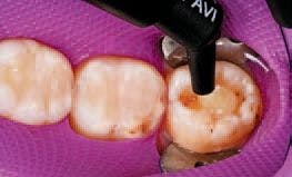

An OptraSculpt sphere was then implemented to sculpt the composite to the floor and walls of the preparation (Fig. 24), after which an OptraSculpt pyramid was used to further form the tooth shape (Fig. 25).
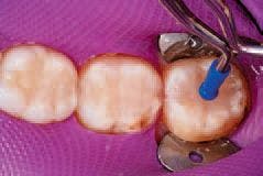

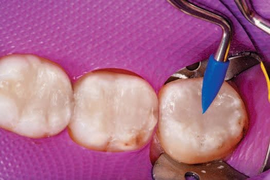
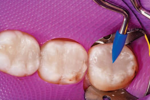
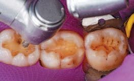
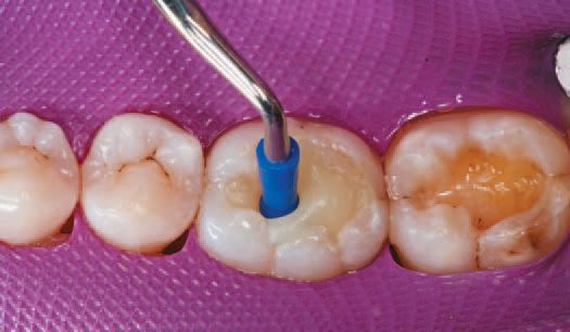
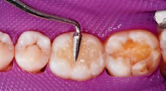
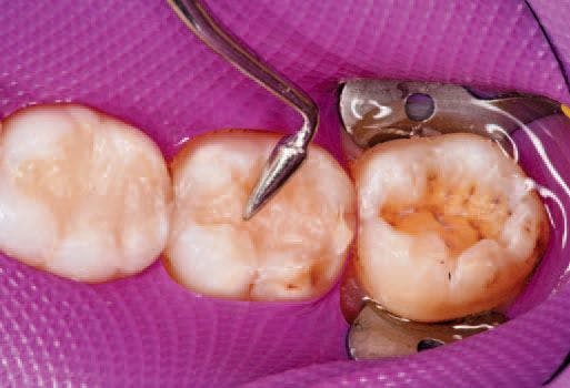
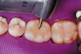
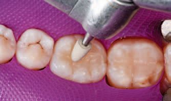
The restoration, now fully formed, was light cured with the Bluephase Style for 10 seconds. Note that the shape of the light’s tip allowed easy access to the tight space. A fine, tree-shaped diamond, similar in shape to the P1 plugger, was then used to establish occlusal anatomy (Fig. 26).


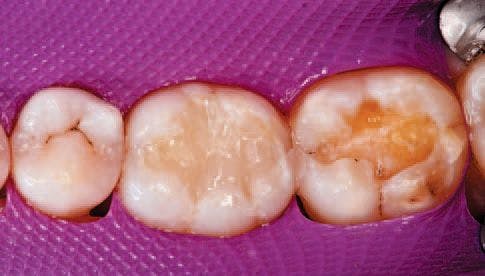
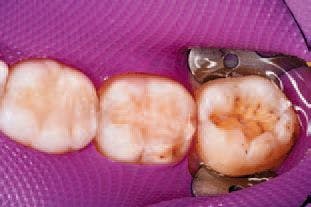
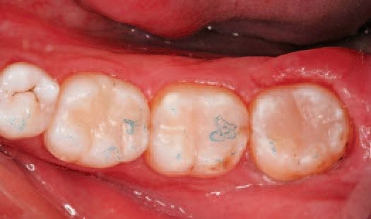
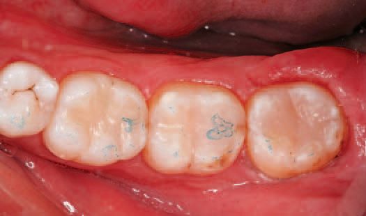
An OptraPol® NG polishing instrument was used to polish the completed restorations of all three teeth to a high-gloss (Fig. 27).


After polishing, the rubber dam was removed, and articulating paper was used to check the patient’s occlusion (Fig. 29).

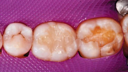
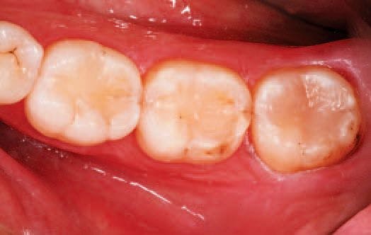
The occlusion was determined to be satisfactory and the restorations complete (Fig. 30). The patient returned the following week for a visit and reported no post-op sensitivity. After re-hydration, the excellent color-matching to the patient’s natural dentition was visible, and all three restorations appeared esthetically similar.


Conclusion
This case illustrates the different reliable methods for posterior restoration placement.
As the materials and processes for posterior restorations have progressed, they have become increasingly more efficient, esthetically pleasing and predictably placed. In this case, the clinician used Tetric EvoFlow and Tetric EvoCeram A3 to place the restoration on tooth No. 31, and the procedure took seven minutes.
The clinician used Tetric EvoCeram Bulk Fill IVA on tooth Nos. 30 and 32, and these restorations were placed in a little more than four minutes each. Tetric EvoCeram Bulk Fill allows for the successful completion of a Class II restoration in half the time.
References on the next page ...
References
1. Giachetti L, Scaminaci-Russo D, Bambi C,
Grandini R. A review of polymerization shrinkage
stress: current techniques for posterior
direct restorations. J Contemp Dent Pract.
2006;7(4):79-88.
2. Cheung GS. Reducing marginal leakage
of posterior composite resin restorations: a
review of clinical techniques. J Prosthet Dent.
1990;63(3):286-288.
3. Ferrcane JL. Resin composite-state of the
art. Dent Mater. 2011;27(1):29-38. Epub Nov
18, 2010. ?


