Intraoral Imaging Is a Team Sport
How the DA, RDH, and DDS can use an intraoral camera to benefit the patient and the practice.


If you follow any team sport, one of the fundamentals of winning is to have everyone on the same page, to know the signals, and to follow the same message. Sure, each position player in any sport has their role, but their main purpose is to collectively steer the team to success.
Practice success comes from great communication. Great communication comes from teamwork. How better to communicate with patients than real-time photos that show, up close and personal, their current health conditions? The adage “A picture is worth a thousand words” is truly evident with the intraoral camera (IOC).
The first intraoral camera, developed in the late 1980s, was an analog IOC system by Fuji Optical Systems of Los Gatos, California. Not only was it cumbersome, but it also came with a price tag of $40,000. Intraoral cameras have evolved from the expensive, analog, floppy-disk, space-hog versions to slim, ergonomic handpieces that have operatory flexibility.
Several features of IOCs include wireless or corded connections, LED lighting, fixed or variable focus lengths, magnification (up to ×100), and different angle views (from 0° to 90°), depending on the manufacturer and style of camera you purchase. All have barrier protection that will not impede the clarity of the digital image.
Many IOCs are portable. Images are immediately available via practice management software and shareable with the entire team. These images enable all members of the team to discuss treatment options and progress. They are also great for documentation and benefit billing and record keeping. Most patients will remember a discussion from 4 weeks ago or 24 months ago while looking at a picture of the area of concern. Images are shareable with other practitioners for an on-the-spot referral.
Additionally, if finances are a shared expense, images may be given to a patient to discuss the necessity for a treatment plan and its financial obligations. It is much easier to show someone a picture of a cracked tooth to explain the need for treatment with their partner than to have them try to describe it on their own.
The Benefits of an IOC
Let’s look at how the IOC will enhance your clinical practice.
The dental hygienists is usually the first clinical team member the patient meets. The IOC is a perfect adjunct to showing areas of concern and teaching proper oral hygiene. One image of the lower lingual arch that shows red, hemorrhagic, gingival tissue with heavy calculus demonstrates a lack of home care to the patient.
For recare appointments, the image is comparable to previous images to show the patient their progress. An image can easily capture areas of heavy plaque accumulation, marginal gingivitis, or bruxism.
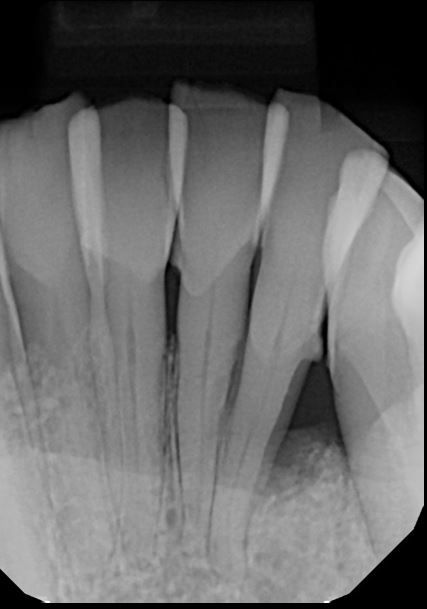
Dental assistants can take intraoral images of a patient’s chief areas of concern prior to the dentist entering the treatment room. For example, a patient of record presents with a fractured lingual cusp. The assistant takes a radiograph of this area, which will offer 1 view of the tooth, whereas the intraoral image will give a clear view of the exact depth of the fracture.
Once the doctor evaluates the tooth, they can explain the necessary treatment, showing the patient both the radiograph (which many patients may not understand) and the clinical image. Dental assistants may use the camera to take initial arch records for the patient’s chart.
Utilizing IOCs is a winning strategy for the dentist. Images will enhance the communication between the doctor and the patient, demonstrate accuracy of treatment areas, and improve treatment acceptance. Images gathered from dental hygienists and dental assistants will facilitate the education of treating patients’ oral health. Early detection of oral health issues is paramount. Treating a smaller, less invasive lesion is more beneficial than waiting until the patient is in pain and a larger expensive treatment is required.
Another benefit is documentation for the future. For example, a patient has been complaining of sharp lower anterior teeth, even after many discussions about bruxism. An image, showing the bruxism, is beneficial not only to planning the oral appliance but also for the benefit documentation. Furthermore, IOC images may be used to document before-and-after treatment.
The business manager’s utilization of the accurate documentation of tooth images is important. More documentation is required for many of the major dental benefit companies. In addition to a digital copy of a radiograph, the digital image of a tooth with a failing historic amalgam, a crack, or an abutment procedure for an implant in place offers visual proof of necessary treatment.
This will assist in obtaining proper coverage and reimbursement for procedures. Additionally, when a patient is sitting with a financial manager and discussing their treatment plan, it is easier to show the patient an image of their treatment plan rather than a group of tooth drawings with colored affects.
Selecting an IOC
So, how do you incorporate the IOC into every team member’s hands? Find a camera that fits into your budget and is available in multiple operatories. For example, the MouthWatch Intraoral Camera starts at $299. Having a few in the office eliminates the need for the team member to go hunting for the camera during treatment.
Some practices have a usage schedule. One would always want a camera available for new and emergency patients, but recare evaluation of periodontal tissues is also important. If fiscally reasonable, consider having multiple cameras available.
The production generated from an extra IOC will pay for the unit after a few services. If the camera is limited to the 1 operatory in the facility, you may be missing out on a plethora of treatment discussions and documentation.
When selecting an intraoral camera, read the reviews and check out the product. It’s important to find a trusted brand that will offer support.
Remember, infection control matters. A barrier sleeve is required to cover the handpiece wand each time an IOC is used. These sleeves are available from the manufacturer, and will accurately fit your wand. Placing the plastic barrier correctly will enhance the clarity of the image.
IOCs are for the whole team. They will enhance communication and trust between the patient and the entire office. Patients give us permission to work inside their mouths. They have little to no idea what is going on or what we see that would necessitate treatment.
IOC Return on Investment
We all have issues with patients agreeing to our treatment plans. Increasing case acceptance is important. By utilizing the IOC, you will realize an immediate return on your investment: Patients will accept treatment more readily.
Showing the patient a lesion or a poor, leaking filling inspires them to want to schedule their appointment. Although we discuss complete periodontal care, some patients have the attitude that “if it doesn’t hurt, it doesn’t need fixing.” With images of the inflammatory tissue changes or bone loss, patients are more likely to complete the scaling and root planing.
When we purchase dental equipment, some items take years to realize their worth. This may be the first piece of equipment that pays for itself in the initial use and is key to case acceptance thanks to the visualization of needs. With an IOC available in every operatory, the message is clearly demonstrated with the images.
Team members will be able to share these pictures with the patient. This will enhance your productivity, encourage referrals, and increase your level of trust with the patient. Having the entire team onboard will be a win-win for the practice. Seeing is believing.