A male patient, age 37 years, presented with failing amalgam restorations with recurrent caries on the upper left premolars (teeth #12 and #13) and first molar (tooth #14) (Figure 1). The preop bitewing was taken to ensure there were no other interproximal caries (Figure 2). All the photos were taken with the CS 1200 intraoral camera from Carestream Dental. After applying anesthetic with 2% lidocaine with 1:100,000 epinephrine, a clamp #24 from HuFriedyGroup was placed on the second molar and a rubber dam inserted.
The existing amalgam restorations were removed using an 806.31.009 inverted cone diamond bur from Brasseler USA. The recurrent caries and amalgam stains were now visible (Figures 3 and 4). All the soft caries were removed using carbide round burs in the sizes RA 2, 4, and 6 with a Midwest Shorty low-speed handpiece from Dentsply Sirona. To make a clean, neat cavity preparation, all superficial caries and stains were removed conservatively with a 6801.31.009 small round diamond bur from Brasseler USA. Intraoral photos were then taken to confirm that no caries were left behind (Figures 5 and 6).
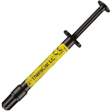
After cavity preparation, the teeth were dried well and a light-cured, resin-modified calcium silicate material (TheraCal LC®; BISCO) was placed on the deep cavity surfaces and light cured using FlashMax P3 curing light (CMS Dental) to protect pulp (Figures 7 and 8). Because TheraCal LC is opaque, it was also used to mask the dark amalgam stains that could show through the translucency of the composite.
TheraCal LC®
TheraCal LC is a light-cured, resin-modified calcium silicate. It is indicated for direct and indirect pulp capping and as a protective liner. TheraCal LC acts as a protectant of the dental pulp and the hydrophilic resin formulation is designed to create a durable liner. It touts moisture tolerance and can be placed under restorative materials and cements.
BISCO
800-247-3368 | bisco.com
After placing Estelite® Sigma Quick composite (Tokuyama Dental) on the lingual of the first molar, Composi-Tight® 3D Fusion Sectional Matrix System sectional matrices and rings from Garrison Dental were placed (Figure 9). Selective enamel etching was then done on the premolars and molar using 37% phosphoric acid (Figure 10). The area was rinsed and dried, and All-Bond Universal® adhesive from BISCO was applied and light cured.
Estelite Sigma Quick composite was then placed first in the second premolar in small increments using a condenser and plastic instrument from HuFriedyGroup. Before light curing the final layer, some occlusal anatomy was created using several hand instruments including the acorn-shaped HuFriedyGroup 21B burnisher, Pioneer Solution Blue Titanium CIB3, and Nordent CEEX3A long explorer (Figure 11). These steps were then repeated on the first molar (Figure 12) and first premolar (Figure 13).
The occlusal adjustments and more detailed anatomy were created using a flame-shaped diamond bur 274.31.016 from Brasseler USA. Finishing and polishing were completed using a large coarse Sof-Lex™ disc from 3M, and a #12 blade and a white Arkansas Stone from Dedeco. The final restorations have a natural esthetic and blend nicely to the tooth (Figure 14). After the restorations were completed, a bitewing x-ray was taken for a final check (Figure 15).