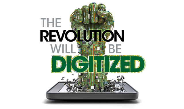
The revolution will be digitized
Digital technology is a groundbreaking force in dental imaging.


Medical radiology traces its origins back to 1895, and it only took two more weeks before Dr. Otto Walkhoff, a dentist from Braunschweig, Germany, applied it to dentistry. Dr. Walkhoff made a dental radiograph using a glass photographic plate wrapped in black rubber and requiring an exposure of 25 minutes. Thus was the first intraoral radiograph.
We’ve come a long way since then, and digital imaging gives us views-of both two- and three-dimensional varieties-that Dr. Walkhoff likely never dreamed of.
The merits of digital imaging
Diagnostically speaking, digital images give faster, clearer, more comprehensive views than their analog predecessors.
“Our diagnostic capability is greatly improved,” Dr. David Rice, DDS, a general dentist in Amherst, N.Y., and founder of IgniteDDS.com, says. “The clarity of today’s sensors is so much better than what we had in the past. Diagnostically, I’m a much better dentist. I can have real-time conversations with specialists, because I can take an image, pull it off my software, email it and in the moment-with the patient in the chair, or vice versa if one of my patients is in their chair-we can be talking back-and-forth about exactly what’s happening as it’s happening.”
Trending article: Why image is everything for dental practices
Digital imaging includes more than just X-rays. For example, DEXIS’s CariVu system uses radiation-free imaging to discover caries in its early stages.
“2D imaging has gone way beyond the X-ray,” Marshall Martin, senior product manager for DEXIS, says. “The X-ray is just one component. There are capabilities for different modalities that are available to actually diagnose people. We’re looking much more comprehensively now. The old idea that you, as a patient, come in, get your X-rays and that’s only what your dentist uses to diagnose is out of date. It’s just waiting until disease progresses to a point that can actually be picked up in an X-ray. In this case, the dentist waits too long and disease progresses, when it could have been found sooner with another modality. One of the things dentists recognize is that if they can detect disease early, the patient may have a better outcome.”
“We’re able to catch decay so much earlier to be more preventive and conservative than ever before,” Dr. Jason Watts, DMD, a general dentist in Cape Coral, Fla., adds. “Everything we do is now enhanced by digital imaging: The root canal that we do and complete; the fillings that we do; the ability to detect decay before it becomes an abscess or infection.”
Doctors are always up against the clock, but when it comes to digital imaging, time is less of an issue.
“With standard analog film, you’ve got to wait for an image to be processed,” Dr. Ed Shellard, D.M.D, vice president of sales and marketing of Carestream Dental, says. “And then you have to determine, ‘Is that a good image or not?’ You may be a few minutes in after you’ve taken the image and then you may realize, ‘Hey, I didn’t get the images that I wanted. I didn’t get the full length of the tooth. I didn’t get the apex,’ and then have to go back and retake the image. But with digital, the image comes up immediately on the screen, and so you can tell right away if you captured the proper area, which is important from a treatment perspective and a time perspective. Within the office, that’s a big help.”
Patient education
It isn’t just the doctor who can see more, thanks to digital imaging. Dr. Rice relies on digital imaging as an important tool for patient education.
“What I can do with digital that I can’t do with plain film is I can change the size of an image so it’s easier for them to read,” Dr. Rice says. “I can modify the image, so I can colorize it, I can do what’s called ‘inverting’ an image, which means we can change how the radiolucency and radiopacity appears on an X-ray, and that just really helps me get a patient on board and understand what I see.”
The ability to communicate that information to the patient aids in treatment plan acceptance.
Martin references a study showing a 25 percent increase in patient acceptance of treatment when digital imaging shows their condition.
More from the author: 9 things you REALLY need to know about digital imaging
“We know that patients are visual learners,” Martin says. “We know when the dentist shares his photographs, images and information in a way that is reasonable for a patient to understand, there’s a significantly higher response for patients. It’s the old adage: If you tell me, I’m going to forget it and I’m not going to care about it; if you show me, I might remember what you’re talking about; but digital allows the dentist to involve the patient, and when people are involved in their own treatment needs, there are better outcomes for the patient and the office.”
The portability of digital images is another benefit, allowing doctors to view patient scans virtually anywhere.
“One of my favorite features of digital imaging is our ability to access images from anywhere,” Dr. Leah Capozzi, DDS, a general dentist in Buffalo, N.Y., says. “I can remote into the system, if I need access to images, to discuss a case with a specialist or refresh my memory about a case. I also like to review the images from my office while the assistant or hygienist is working on the patient. It makes it so much easier to start treatment planning and get my thoughts organized prior to sitting down with the patient.”
“We’re seeing more and more offices and practitioners combining offices and going into group practices, or even medical facilities,” Jan Myskowski, clinical program manager, 2D and Small Equipment at Kavo Kerr Imaging, adds. “The doctors travel from office to office, and they’re needing the use of the application devices, so if they have a smartphone or if they have a tablet, they want an application where they can view these images and diagnose remotely.”
Up next: Benefits of 3D imaging ...
The third dimension
Two-dimensional images are certainly useful, but in recent years, three-dimensional, cone beam computed tomography (CBCT) has made it possible to see even more.
“3D brings the third dimension,” says Peter Soto, 3D systems product specialist at KaVo Kerr. “2D is flat. It doesn’t allow the doctor to see what’s really going on. 3D changes all that. It’s like a GPS for the mouth. You can go further into the patient’s anatomy and see what’s really going on. You can see it, diagnose it and treat it with much greater accuracy. It allows the clinician to see what he couldn’t see before and it allows him to be more prepared than he could have been before.”
“When you get to 3D, that changes the whole picture,” Dr. Jay Reznick, DMD, MD, director of the Southern California Center for Oral and Facial Surgery, in Tarzana, Calif., and founder of Onlineoralsurgery.com, says. “Life is in 3D. Patients are not two-dimensional creatures. They’ve got anatomy, they’ve got different structures around the jaws and when you’re looking at a two-dimensional image, you get superimposition of multiple anatomical structures on top of each other, and sometimes it’s very difficult to discern what’s what. So you may see things that look like they are pathologic, but they aren’t really anything to worry about. You also miss things that are significant pathology because you can’t see them. For example, it might be hidden behind a root or bone density.”
Related reading: How to integrate dental digital imaging with your paperless workflow
The abilities of 3D imaging go beyond conventional dentistry, extending into additional areas beneficial to patients.
“Dentists are starting to ask their patients, ‘How are you sleeping?’” Kenna Wuistingher, 3D clinical program manager at Kavo Kerr Imaging, says. “They know that sleep affects your overall health. They are not just general dentists anymore. They’re going to the next level, and they’re treating overall health and educating more, and that comes from knowing what your airway analysis reveals about sleeping, breathing and how these factors affects a patient’s day-to-day function.”
Dr. Reznick doesn’t always use 3D imaging, but it has become more commonplace.
“Sometimes a two-dimensional image doesn’t always give you the full picture,” Dr. Reznick says. “For example, for wisdom teeth, we used to just get a two-dimensional panoramic, because that’s sort of what was recommended by The American Association of Oral and Maxillofacial Surgeons (AAMOS). What we were finding was, at least half the time, that there was concern regarding positioning of the teeth or position of the mandibular nerve or the sinus, so we would get a cone beam scan, in addition to a panoramic, to get that additional anatomic information. And what I realized was that a good majority of the patients that we do the cone beam scan on would have been missed if we had just done a two-dimensional scan. So now I tend to do a three-dimensional, a lot more commonly.
“The only thing that the cone beam isn’t that good for is looking at caries,” Dr. Reznick observes, “because if the patient has a lot of crown and bridge or restorations of their mouth, that causes scatter, and that scatter makes it difficult to interpret the image.”
Safety
Radiation exposure is a real concern.
Not long after Dr. Walkhoff pioneered dental radiology, he and a gentleman named Fritz Giesel established the world’s first dental roentgenological laboratory. Giesel died a year later of metastatic carcinoma caused by heavy radiation exposure to his hands.
While the safety of conventional radiology has improved since the 19th century, digital imaging makes it is even safer.
“With 2D intraoral imaging, it’s lower radiation,” Myskowski says. “Of course we want to provide a good service for our patients and our staff, working with radiation to limit that exposure. So manufacturers are able to go lower on our exposures utilizing sensors and/or phosphor plates, and the technology of the software, in conjunction with the imaging and the lower radiation, we can get an optimal image and be able to diagnose off of this so much better than we had in the past with either film or other venues that we were doing.”
“Digital imaging is much safer than conventional imaging,” Sean Mitchell, imaging specialist at ACTEON North America, says. “The amount of radiation needed to produce a high-quality image is approximately 80 percent less than film. This radiation savings is very favorable for the patient; however, it is even more critical for the office staff responsible for acquiring the X-ray images. Also, when an office moves to a digital format it eliminates the need for costly, harmful chemicals that are required for developing X-rays.”
In addition to technology lessening the amount of radiation patients and staff are exposed to, improved imaging quality results in even fewer doses.
“Dosage is something that gets brought up all around the country from our patients and our doctors,” Wuistingher says. “We want to better serve our patients, so overall better health of the patient is the biggest criteria. By offering 3D imaging and 2D imaging, compared to the film we were using before at a higher dosage, the image quality has improved so much, plus you’re able to adjust and change filters and enhance that image even more. You really don’t take a bad image anymore. You’re not reradiating a patient because you got a poor image. You can almost guarantee you’re going to get a great image every single time.”
Software
While the hardware is responsible for the core operation of a scanner’s duties, software is necessary to view and interpret the data.
Interesting read: 5 ways digital imaging improves the patient experience
“Software is imperative to the process of capturing and storing digital images,” Mitchell says. “Imaging software applications apply custom developed filters to each digital image in order to enhance them. In most cases, imaging software will offer multiple filters in order to customize enhancements, per a clinician’s preference. SOPRO Imaging by ACTEON provides an office with three customizable filters to choose from. This ability to manipulate the images reduces the need to retake images and enhances the ability for diagnosis.”
Software allows doctors and other staff members to personalize and adapt the image results to their own preferences. The result helps clinicians better interpret the results.
“We have a product called CS Adapt, which actually allows the practitioner to set up, within the software, the capability to set the image based upon the practitioner’s desires and look and feel of the image,” Dr. Shellard says. “Within imaging, as a whole, there’s a lot of subjectivity. Some docs like a sharper image, some like a less sharper image, some like a different type of grayscale. What we’ve done is tried to present the image in a way that they’re used to seeing it. Let’s say they’re used to using Insight film, or a standard analog film, and they like to see the image that way. One of the biggest hurdles for docs, as they transition from analog to digital, is they don’t necessarily like the digital image. And that is not necessarily because of the precision of the image, it’s more of the subjective nature of it showing the doc that it doesn’t look the same. So while having these preset looks, the doc can actually capture the image and look at it the way they want to see it.
“It also holds true within the practice,” Dr. Shellard continues. “If you’re a hygienist and you want to see an image a certain way, because you’re more concerned about bone height and potential periodontal issues, you can have a periodontal filter-or if you’re an endodontist and you want to have an endodontic look. So the ability to personalize an image, depending on the situation and depending on the objective needs of the practitioner, becomes really important.”
Sometimes the technology behind the software comes from unique sources of inspiration.
“We also have another product called Logicon,” Dr. Shellard says. “It’s a software that actually piggybacks off of the digital image and it applies algorithms to the interproximal areas of the teeth to determine the extent of the decay. It was, ironically, built as an outgrowth of the work done in the aeronautical industry. So if you can imagine that you’re looking at satellite photos and you’re trying to determine if there are tanks or missiles. As crazy as it sounds, Logicon was developed by a rocket scientist.”
Software also allows scans to be processed differently for unique applications. So rather than repeatedly scanning the patient, that initial scan can be reinterpreted.
“Dentists are considering overall health,” Wuistingher says. “They are not being reactive; they’re proactive. They are taking an image at a lower dose and now have so much more data to read. They are providing airway [TMJ] analysis and planning implants. They are seeing images in full 3D, in cross-sections and from all directions. And dentists are doing this from one captured image that’s available in the operatory within a minute.
“There could be a fractured tooth or restoration that can’t be seen by looking in the mouth or felt by the patient, but when the doctor takes this type of image, he or she can spot the condition before it becomes a bigger issue for the patient and they’re in pain,” she continues. “There’s a lot of early detection that can be done now.”
Up next: The advantages of digital imaging ...
Benefits
Everyone can get something out of digital imaging-whether it be in 2D or 3D.
“It’s really something that can be used in any type of dental practice,” Dr. Reznick says. “Obviously, if you’re a surgeon or a surgical specialist you’re using it for looking at pathology, for planning implants, when looking at the temporomandibular joint. If you’re on endodontist, you’re looking at the periapical regions. If you’re a periodontist, you’re looking at the bone level around the teeth. But if you’re a general dentist, you can obviously look at those same things. In addition, it’s going to show you, possibly, early pathology that you would be scratching your head not knowing what’s going on. So you can pick up pathology much earlier in your practice. If you’re planning implants, obviously it’s very beneficial to plan in 3D and know what, anatomically, the limitations are.”
“If you look at it from a pathology standpoint, I can visualize it so much more,” Dr. Rice says. “I want to keep my patients safe, so I love that. If I’m a dentist who does root canals, if I’m doing endodontics, the number one failure for root canals is because we’ve missed canals. So if I have a 3D image, I’m not going to miss anything anymore because I can see all those tiny little canals that I couldn’t see before, and then it opens up so many opportunities, so people who are looking at airways and sleep with dentistry, that’s awesome. People who are placing their own implants, it revolutionizes that. And then the pinnacle of it is the 3D image and CAD/CAM. That integration piece is massive. There’re so many avenues for today’s practices to bring 3D onboard.”
Trending article: How is dental technology REALLY being used?
CBCT imaging might not be for everyone, but certainly specialists can benefit from it.
“3D imaging can be costly, so it depends on the work being done in the practice,” Dr. Capozzi says. “I think that excellent results in endodontics and implant dentistry can be achieved using 3D imaging. If a general dentist is placing his or her own implants, 3D imaging should be considered. As far as specialists go, I would certainly be more likely to send a patient to a surgeon or endodontist who is using 3D imaging over one who is not. A great benefit of 3D imaging is guided implant surgery. This allows us to virtually plan the implant placement based on the position of the restoration and quality of the bone. You can fabricate a surgical guide and assure that the implant is in the exact position you need.”
While CBCT systems can be expensive, dental manufacturers have taken steps to make the prospect a little more palatable.
“We have systems that let the doctors upgrade,” Soto says. “It allows practices to grow around their system. They can bring in a smaller field of view at an economical price point, and then upgrade their system with much larger FOVs as they evolve. Or they can jump right to the highest level. The doctor can control where he or she wants to go next with the next size and yet retain [the] hardware and software.”
Why doesn’t everyone have it?
So why isn’t digital imaging in every practice everywhere?
While it’s easy to blame costs (and that’s the big one), doctors’ willingness to upgrade is an important factor.
“Digital imaging is a faster, more accurate and cost-effective way to capture images than film; however, not all dental professionals have adopted the new technology,” Mitchell says. “It is estimated that 20 to 30 percent of dental offices still use film to acquire X-ray images. One reason for this is that dentists have conventional radiography devices in their offices that are still functioning. Converting to digital imaging can be perceived as expensive. Also, the transition from conventional to digital radiography requires training for the entire staff. Not only may there be placement training needed, but also time to learn how to navigate through the imaging software. An office may not want to incur any downtime due to training their team on the new imaging system and its processes.”
While change can be a difficult hurdle to clear, Dr. Watts looks for new dentists to help usher in that change.
“Change is scary, and something that will cost you $20,000 or $30,000 is a scary thing when you’re going to retire in a few years,” Dr. Watts says. “But a new generation of dentists don’t know anything but digital radiology. If people just overcame the fear, then they would know that they would be treating and servicing patients better in every way, shape and form. And they’ll make up for it, because they’ll reduce the time the patient’s in the chair, and they can see more patients. I don’t think people are processing that. They just look at the sticker price.”
Something as simple as the doctor’s own comfort zone is another factor.
More from the author: What dentists want labs to know
“For some docs it’s cost, for others they like the quality of film and they don’t want to switch,” Dr. Shellard says. “And you see that in the everyday world too, where you have these audiophiles that will never give up their vinyl records. They won’t go digital on music, because they think the sound quality will diminish when you go to digital. There are a group of folks who will probably never give up analog film, because they love that quality.”
Even though imaging prices can be off-putting, the ability to upgrade machines is an option. For instance, Wuistingher says that some of their machines can be expanded as the practice evolves.
“Even if you’re the general practitioner that is starting out, coming straight out of dental school, you can purchase a product that fits your needs now,” Wuistingher says. “And if you decide to add airway and TMD treatment, larger implant cases or ortho-any new area where you need more anatomical information-you can always upgrade those products.”
In the 19th century, Dr. Walkhoff made an immeasurable impact on dentistry. And in the 21st century, doctors have even more amazing tools that allow them to deliver better care.
“Digital imagery isn’t the future, it is the now,” Dr. Watts says. “Soon it’ll be the standard. Those who are waiting are really procrastinating, because sooner or later every office will have it.”


