Why image is everything for dental practices
Dr. Flucke explores how communicating with images-and having imaging systems that communicate with one another-can make a big difference for your practice and your patients.


I've just finished reading a few articles written by attendees at the 2017 Consumer Electronics Show (CES) in Las Vegas and one point continues to be reinforced by every tech book and article I read. Simply put, our practices and our world continue to evolve in a more and more digital manner.
Nowadays, the majority of things you want to add to your practice is going to either require a computer connection or will somehow create digital data. While anything digital is going to be faster and more efficient, there are some downsides to current digital systems. While it pains me greatly to type those words, I have to admit that they are, in a way true. The main drawback to which I’m referring is the current lack of communication between many of our systems.
Can’t we all just get along?
In the early days of technology in dentistry we went through what I refer to as the “Innovation Phase”. This phase saw the creation of several technologies such as intraoral cameras and digital radiography. While these, and other, technological wonders began to make inroads and change the way we practiced, they were standalone units that were kept on carts and brought into the treatment area only when they were needed. This did, of course, slow down the adoption of these new technologies but despite these shortcomings, people saw the advantage of visual communication.
What really changed things and fueled a rapid growth in technology adoption in dentistry was the revolution in computer processing power. As computers became more and more affordable, they began to migrate into the operatory. This was driven by low cost and the fact that practice management software had begun to include clinical packages that allowed for charting… and most importantly, imaging.
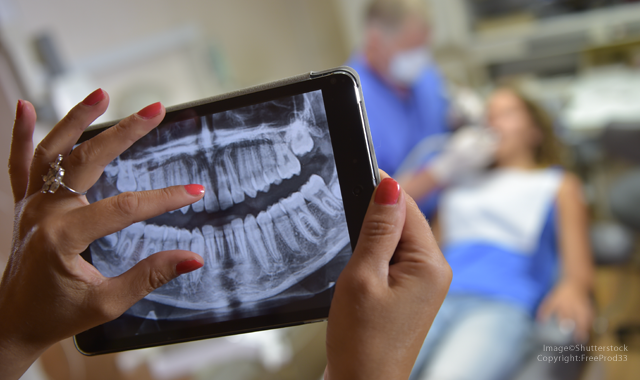
For manufacturers this meant they could do away with much of the clunky hardware that made their products cart based and connect them to the operatory computer instead. Suddenly the intraoral camera and the digital X-ray sensor could connect to the computer, be shown to patients on the screen, and stored directly into the patient’s digital chart.
What followed was more and more technologies began to be connected to the operatory computer. This made the computer the technological hub and the devices were all spokes that connected to it. This is what I refer to as the “Integration Phase”.
We progressed fairly well with integration until around 2007. At that point the profession began to see growth in two fields: digital impressions and cone beam 3D radiography. These two technologies were standalone, using their own computers and their own software. The standalone aspect of these technologies worked well at first. Cone beam was very expensive in the beginning and digital impression systems required doctors to revamp their approach to prosthetics. These factors kept the growth of the technologies to modest levels.
Yet, like what happens with any successful product, the more it proves itself successful the more users being to adopt it. Currently we are seeing both of these product categories passing the realm of the early adopters and moving more toward mainstream adoption. Because of this, dentistry is now in need of another “Integration Phase”.
Picking gnats out of pepper
As someone who has been using an i-Cat cone beam and the iTero digital impression system for over seven years, I’ve had the opportunity to see our practice grow in usage and understanding of these unique technologies. While our early days of usage didn’t require universal access to the data, we are now at a point where I would prefer one software where we could store and access all our imaging files.
That doesn’t mean I’m opposed to sophisticated software to edit, treatment, plan, etc. It would just be nice to have a basic viewer built into my management software as well as a universal place to store this data and back it up. As things currently exist, if an office has multiple imaging modalities at their disposal, chances are those differing modalities are stored in different locations. That includes offices that use a digital radiography system that is independent of the practice software. Backing them up successfully and regularly requires a degree of tech expertise that is not always readily available in the normal dental office. This doesn’t even factor in the possibility of an office adding a technology that they don’t even know requires backup.
Communication breakdown
Our profession sadly lacks integration (See? There’s that word again.) between the software that runs our practices and the digital sensors that provide our radiographs. This lack of total integration has been going on way too long in this industry.
For years now, I’ve had people call or email me wanting to know my opinions on a certain brand of digital X-ray sensors. To their surprise, my first question is almost always, “what practice software are you currently using?” The reason for that is because some sensors work very well with some practice software programs and some do not. If a doctor is happy with their software, I encourage them to explore the sensors that work well with that software.
My reason for that is efficiency both in clinical situations as well as running backups. Well-integrated sensors usually store their images in the software’s database meaning you only have to back up one system and there are many fewer clicks when navigating the images.
An office can certainly run whatever sensor they like, but that can require a separate viewing software and backup which complicates matters.
In the world we live in now, there is no reason that almost all sensors cannot communicate with any program. The current situation shows either laziness on the part of the companies involved or a deliberate effort by the companies to limit your choices. Either way, it needs to change.
Digital radiography is now in about 50 percent of dental offices. The time for open source and choices for the doctor is long overdue.
Say it with pictures
In addition to computer prices dropping, we’ve also seen a stunning decrease in the price of hard drives and flat screens. That means there really isn’t an excuse anymore when it comes to taking pictures and sharing them with patients.
When setting up for a procedure, I highly recommend the staff always makes sure the intraoral camera is ready. Everyone on my team that has clinical responsibilities is well versed in using the intraoral camera. Whether it is taking pre-op pictures before we start a procedure or photos to help explain diagnosis and treatment to a new emergency patient, the staff uses our cameras frequently. Hygienists take lots of photos to help educate and to help in the co-diagnosis phase when I need to explain treatment needed.
The same applies to our visually based caries detection devices. Human beings are visual animals and experts routinely tell us that around 85 percent of what we perceive about our environment is delivered to our brains via the visual sense. Utilizing a bit of logic here, why would someone choose not to engage the sense that provides 85 percent of our information? Reversing that logic, without pictures patients are operating on an input system that delivers 15 percent of what we are trying to convey. That makes your camera and patient viewing screen seem pretty important, doesn’t it?
We also utilize DSLR cameras in our practice and the team is well trained in how to use them. A photograph taken with a high-resolution camera can impart lots of info, not only to the patient, but to the doctor as well. I routinely consult these photos when treatment planning cases.
When seating cosmetic restorations, we have the pre-op photos on the patient monitor to help the patient remember what their appearance was before we started. This really helps when they see their new smile for the first time and can compare it with an enlarged photo of what they looked like preoperatively.
Wrapping up
To conclude this month’s Technology Evangelist column, I’d like to encourage doctors to take more pictures! Through greater utilization of technologies that you already have in the practice you can make you treatment as well as patient communication easier.
To the manufacturers, I’d like to encourage you to solve the problems regarding choice and open source that the profession is currently facing. Everything goes better when we all work together


