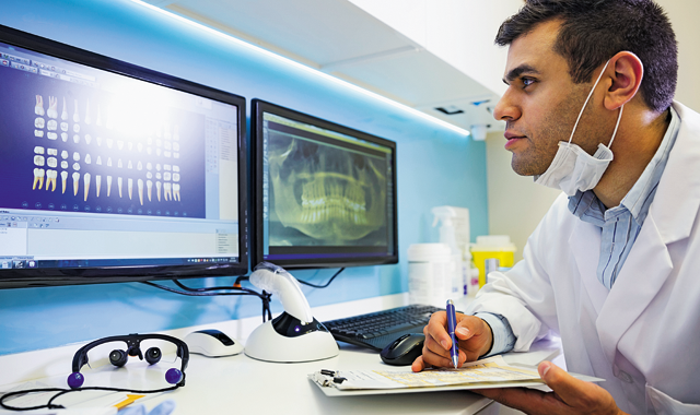
How to integrate dental digital imaging with your paperless workflow
The primary benefit of using digital images is that they are–wait for it–digital! It seems obvious and yet it is often overlooked. So, what makes digital images so special?


The primary benefit of using digital images is that they are–wait for it–digital! It seems obvious and yet it is often overlooked. So, what makes digital images so special?
At the basic level you need to realize the terms “paperless” and “digital” are interchangeable. A paperless chart is a digital chart and a digital radiograph is a paperless radiograph. It is not possible to have paperless records unless you have paperless diagnostics such as digital radiographs.
Trending article: The importance of becoming a digital dentist
In the most literal sense, to digitize something means to turn it into digits or numbers. In a more practical sense, it means turning something into the electronic language a computer can understand. Digital data-everything from words and numbers to pictures, radiographs, sound and video-can be stored, transmitted and enhanced electronically with a computer and a computer network. The concept is simple but the impact is enormous.
Trending article: How to ensure the entire team is on board with technology
Continue to page two for more...
Speed
The most obvious benefit of digital images is the speed of acquisition. You can view a digital photo or a digital radiograph seconds after exposure. Compare that to waiting five to 10 minutes for a film radiograph to be processed or several days for a film photo to be sent off, processed, printed and returned.
Trending article: Top technologies for caries and cancer detection
Clinical results
If you take a radiograph that is clinically unacceptable-you missed the apex or overlapped the interproximal-with film you won’t know for minutes. Once discovered, you’ll need to start from the beginning with film placement and cone position and hope you get it right the second time. With a digital image, you can see results in seconds, and the sensor and cone will still be in place. If you missed the shot with , you can immediately re-position for a perfect image in seconds.
Trending article: 5 technologies your practice needs to invest in now
Continue to page three for more...
Enhancement
Enhancement means we can apply software tools to improve diagnostic efficiency. The most basic tool is “zoom”-blow up the image to see details more clearly. This helps the dentist diagnose and allows dentists to show patients key areas for education and treatment presentations.
More advanced tools allow us to change contrast brightness and other features of the image to enhance diagnosis in ways that are simply not possible with film. For example, as dentists we use radiographs to diagnose several different disease types, caries, bone loss and periapical lesions.
We are looking at several different tissues-bone, pulp and tooth among others. There is an optimized way to image each tissue and to enhance the detection of each disease type. With film we have one (rather tiny) static image we need to interpret for all these things. With digital we have an image that can be enlarged and enhanced in different ways to give us optimized views for each tissue and each disease.
One of the most common mistakes dentists make with digital radiographs is to compare a simple raw digital image with film and decide whether digital looks like film. We are still in the middle of a difficult shift in diagnostic practices and much of what we do with a digital image is tied to what we used to do with film.
The fact is, the digital image contains a lot of data we simply cannot see unless we use dental imaging software tools to enhance diagnosis. Then, we can see things that would never be apparent on film.
This is an area that still needs a lot of research and development. That is, what does the data on a digital radiograph tell me? What is the optimal image setting to detect caries or bone loss? Which tools are diagnostic and why? And what features did we look for in film, which are simply no longer important with enhanced digital images?
Continue to page four for more...
Storage and transmission
Storage just means a digital radiograph can be stored on a computer hard drive with no paper folders. A more advanced feature of storage is to store the radiograph away from your office in the cloud. Cloud storage gives you greater security and easier transmission.
Transmission means the digital data, the “stuff,” can be sent anywhere in the world using a computer network. That includes your office network, which means we can view a radiograph at various places in the office-anywhere there is a computer. You also can connect to the biggest network of all, the Internet.
Trending article: The state of digital radiography in the dental practice
We can send the image to an insurer for payment, a surgeon for referred treatment or to a radiographic specialist for diagnosis. When we send a radiograph to a third party we still have the original and the copy is every bit as good as the original. With film we either lose the original or send a copy that is always significantly less diagnostic than the original.
We have seen the future and the future is digital. The future is coming and it will be amazing!


