The reinvention of dental imaging software
Exploring new technologies that will enhance practice imaging capabilities.


You may not know the name, but if you’ve read a dental radiograph, it’s even money that you’ve used a product invented by Kevin Crucs. I recently had the opportunity to talk with Kevin, the founder and VP of R&D at Apteryx, to explore the rapidly evolving dental imaging landscape.
You didn’t set out to build a dental imaging company. How did it happen?
It was 1995 and I had started a custom software company. Our major client had just released their first product, so we had some downtime while they developed the next set of changes.
My father, a dentist, was concerned with insurance companies destroying his radiographs, which, at that time, required him to submit the original X-rays with the claim. During my company’s downtime period, my father asked us to write a program to digitize conventional film, allowing for electronic transmission of copies to the insurance companies and other specialists. The insurance companies took notice and especially liked our format for saving and archiving the images in an unmodifiable format, which we patented. Later, equipment vendors asked us to modify our software for digital sensors, and we’ve spent the last 20 years refining our imaging products around that core technology.
More from the author: How to solve the peri-implantitis problem
What kind of trends are you seeing in the dental imaging arena?
For one thing, the role of hardware and software in the choice of dental imaging systems is shifting. Traditionally, the imaging platform decision has been hardware-driven. The strategy of many imaging vendors has been to provide “free” proprietary software that locks buyers to their equipment platform and future hardware upgrades. Other areas of medicine have insisted that equipment vendors adhere to open standards like the DICOM format and protocols. In contrast, dentistry has evolved into a fragmented landscape of hardware platforms that don’t work well together.
The costs of imaging equipment, including digital sensors, intraoral scanners and even CBCT, is coming down fast. When digital sensors were an emerging technology 10 years ago, it was “Doctor, you can buy a couple of digital sensors for your office or buy yourself a brand-new BMW.” Today you can get very capable digital sensors for a few thousand dollars. However, mixing and matching new equipment is complicated by closed legacy systems and proprietary data formats - especially when considering integration with practice management and clinical charting systems. As a result, there’s a much greater awareness of the importance of software in the dental practice imaging platform decision process.
What do you see for near-term innovation in dental imaging?
Even though it’s harder for imaging companies to separate themselves with equipment, there will be a lot of innovation, enhanced functionality and diagnostic tools coming from the software side. We’re on the verge of some significant advances in the areas of open integration, migration to the web and artificial intelligence applied to imaging. While there has been a steady evolution of dental imaging in the last 20 years, the pace of change is going to accelerate with multiple major innovations that will come in the next few years.
I recently did a column on artificial intelligence (AI) for caries detection. Is that how you envision AI being used in imaging?


Caries detection is certainly an area in which we’ll see beneficial AI enhanced imaging software in the next year or two. There are several true AI applications for aiding diagnosis and doing other types of feature identification on images that are approaching commercial readiness.
We’re also very enthusiastic about incorporating AI into our imaging software from a clinical quality assurance standpoint. We foresee software that will help the dental office get good X-rays by using AI algorithms to analyze the image database and generate reports that highlight image quality issues and image acquisition issues. Let’s say a user has 50 percent more cone cuts than average. That might point to the need for additional training on positioning. If the X-ray generator is producing substandard images, then perhaps the settings need to be adjusted - or it may be time to buy new equipment. A range of quality assurance measures can be enabled with AI, which systematically analyzes huge image libraries for problems, associates them with potential causes and provides potential solutions.
Trending article: Why cloud software isn't the future
You mentioned migration to web platforms as a coming trend. That migration has been slow in coming in the practice management space. What’s the situation in imaging?
There’s no question that migration to the web should be considered by practices of all sizes and types. The initial impetus is reducing costs such as purchasing local servers, IT support, maintenance contracts and time-consuming data backups. DSOs and large group practices are already moving to cloud-based imaging solutions due to the magnitude of these costs at scale and the need for platform standardization, data portability and central reporting across locations. Private practice owners often wind up doing a lot of DIY tech support. So in addition to cost benefits, moving to a cloud-based imaging system removes a source of frustration and distraction.
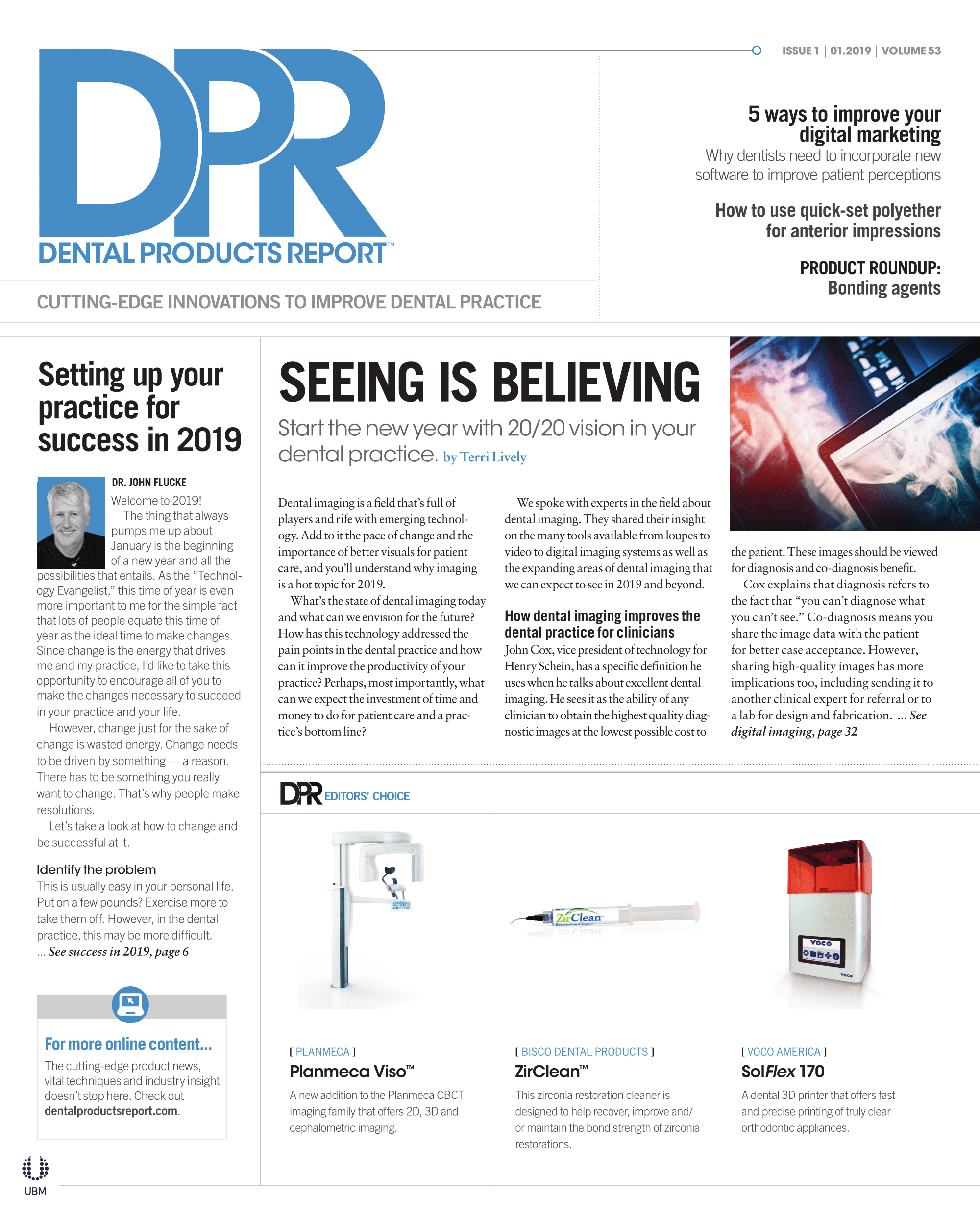
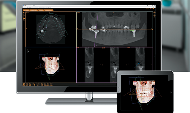
As importantly, migrating to the cloud facilitates gathering important practice data in one easily managed environment. A typical practice may start off with a 2D X-ray system. They later add an impression scanning system and then a cone beam. The vision is to overlay the 2D and 3D images for comprehensive diagnosis, treatment planning and appliance fabrication. Moving to a cloud-based platform makes it possible for us to provide the desired user experience and help dentistry realize the combined promise of these powerful imaging technologies.