How to detect caries around amalgam restorations
How the Canary System helps solves a common clinical challenge by detecting caries around the margins of amalgam restorations.


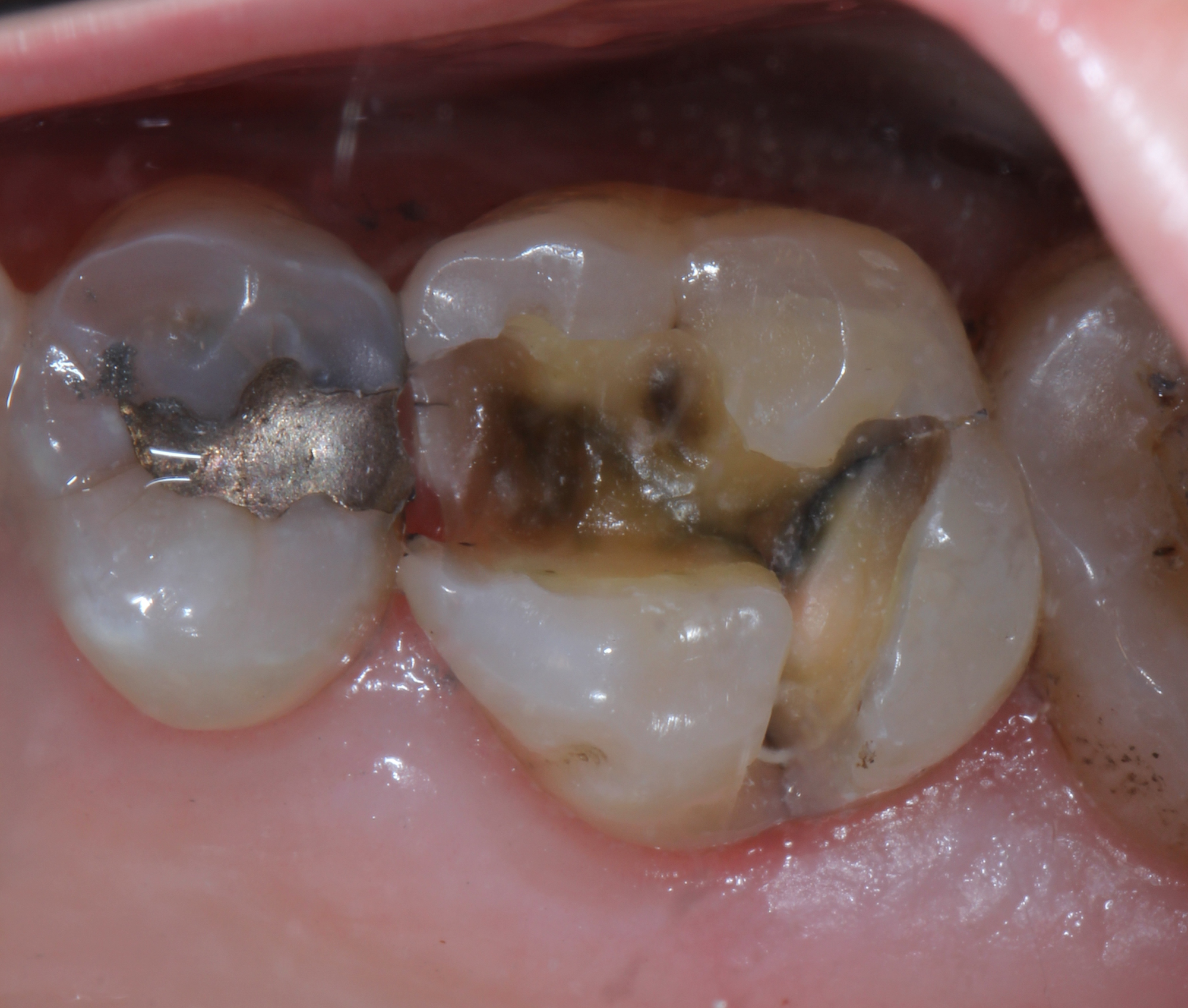
Detecting caries around amalgam restorations is a major clinical challenge. Typically, older amalgam restorations may cause some marginal staining, but visually the margins may appear intact and sound.
The detection of secondary caries in its early stages is not easy.1 Using detection methods such as radiography, explorer, fluorescence caries detection aids and visual examination may not provide any relevant information. Discoloration next to the restoration or ditched amalgam margins is not necessarily predictive of secondary caries.2


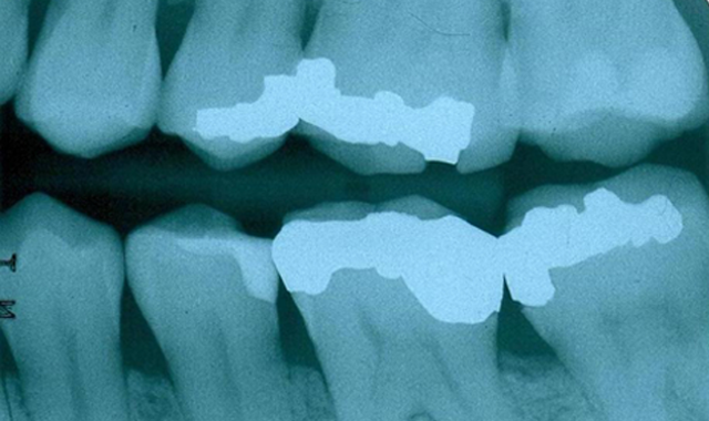
Fig. 1
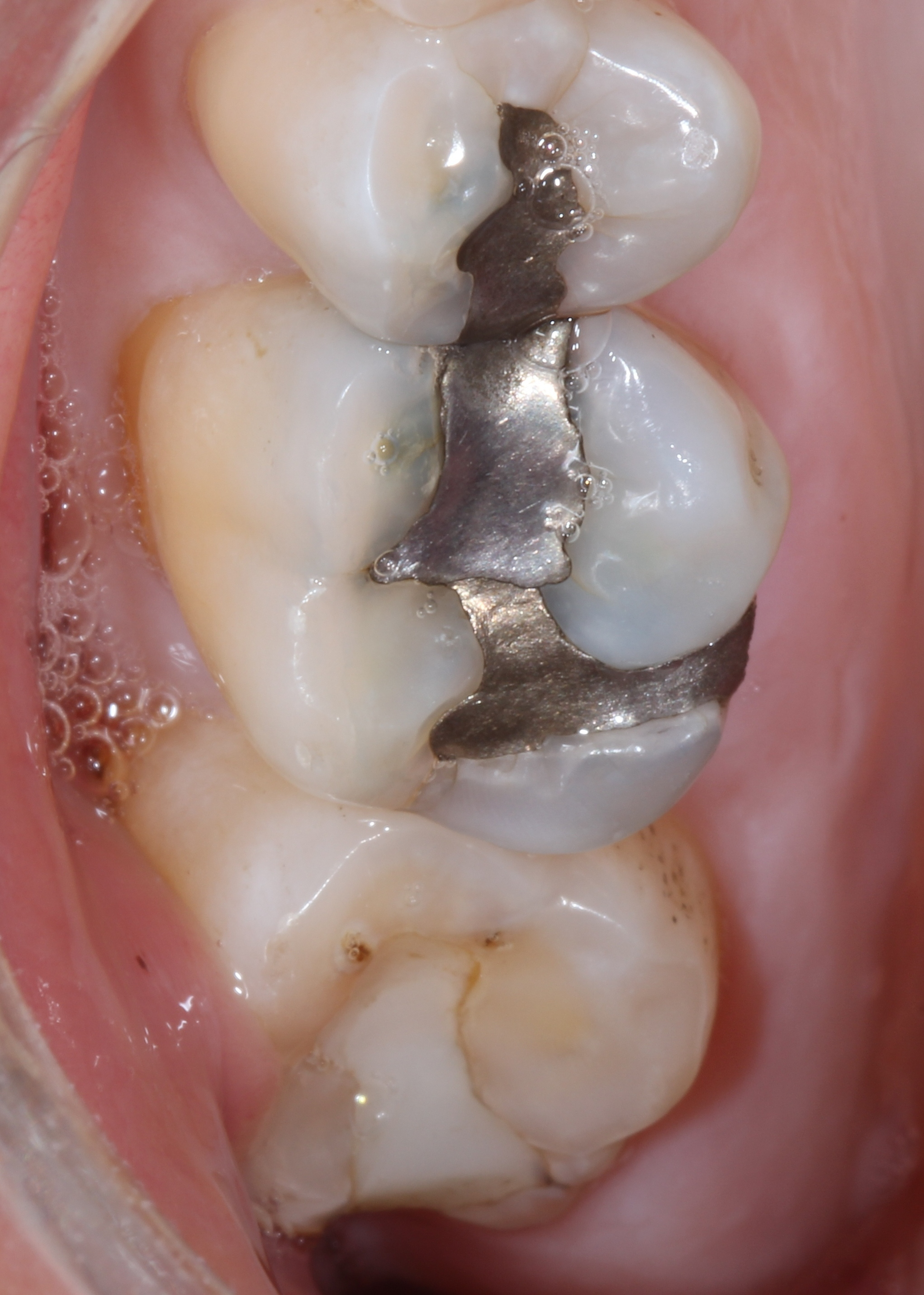
In this clinical situation, the patient had been complaining of pain on the maxillary left side. Visually, the restorations appeared worn but the margins were intact (Fig. 1). A bitewing radiography did not reveal any radiolucency around the restorations on the teeth (Fig. 2).
Bitewing radiographs may not be the ideal for detecting enamel secondary caries,3, 4, 5 particularly if they are located on the occlusal or smooth surfaces. The restoration also masks the ability of radiographs to detect caries along the preparation walls.6 All of these factors create difficulties in determining the source of pain.
Trending article: The top 5 how-to techniques of 2016
We were faced with a challenge. Do we watch and wait for further symptoms to develop? Do we remove the restoration to see if we can detect caries? Are there other caries-detection devices that can detect caries around the amalgam margins?
There are a number of caries detection devices on the market today, but they have limitations. The fluorescence-based devices such as SPECTRA, DIAGNODent and SOPRO are not detecting caries, but other components that glow or fluoresce at a particular wavelength. The literature indicates that: bacterial porphyrins (bacterial breakdown products), stain, tartar and food debris all fluoresce under the wavelengths used in these devices, whether or not caries is present.7 8 9


Fig. 2
A number of studies have concluded that measuring fluorescence is not suitable for detecting caries around restoration margins or beneath dental sealants due to false positive readings.10, 11, 12, 13 The CR Clinicians’ Report (March 2012) found that existing restorations interfered with readings.14 Fluorescence does not give any information about lesion size or depth, and does not penetrate beneath the tooth surface due to the scattering of light from stain, plaque, organic deposits and surface features such as pits and fissures.15, 16
Continue to page two for more...
The Canary System (Fig. 3) uses energy conversion technology (PTR-LUM) to image and examine the tooth. Pulses of laser light are aimed at the tooth, and the light is then converted to heat (Photothermal Radiometry or PTR) and light (luminescence or LUM), which are emitted from the tooth surface between pulses. These harmless pulses of laser light enable the clinician to examine sub-surface caries up to 5 mm below the surface.17, 18


Fig. 3
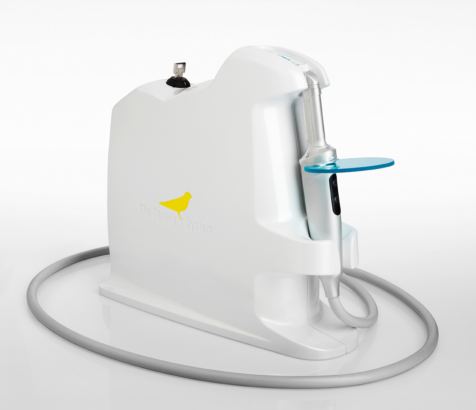
Caries modify the thermal properties (PTR) and glow (LUM) of healthy teeth. As a lesion grows, there is a corresponding change in the signal. In effect, the heat confined to the region with crystalline disintegration (dental caries) increases the PTR and decreases the LUM. As remineralization progresses and enamel prisms start to reform their structure, the thermal and luminescence properties begin to revert towards those of healthy tooth structure.19, 20, 21
Trending article: How to treat proximal caries lesions
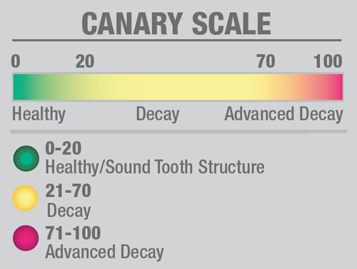
The Canary System creates a Canary Number (ranging from zero to 100) from an algorithm combining the PTR and LUM readings, which are directly linked to the status of the enamel, dentin or cementum crystal structure22 (Fig 4). A Canary Number of less than 20 indicates healthy crystal structure. A Canary Number greater than 70 indicates a large lesion that may justify restoration. Canary Numbers falling between 20 and 70 indicate the presence of early carious lesions or cracks that may require restoration, particularly at restoration margins.23 If the caries is located beneath a healthy layer of enamel, The Canary measures both healthy tissue and caries. The healthy crystal structure overlying the caries dampens the signal, decreasing The Canary Number.


Fig. 4
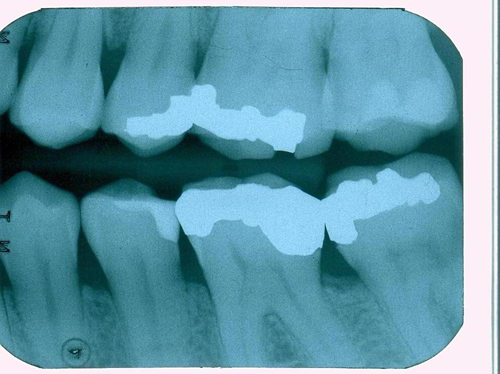
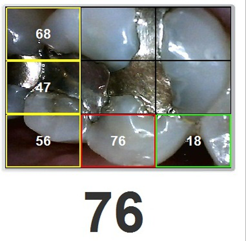
We scanned the margins of the maxillary first molar with The Canary System and found Canary Numbers ranging from 47 to 76 around the margins of the amalgam (Fig. 5). Removal of the amalgam restoration confirmed the presence of caries in both areas (Fig. 6).
A study presented in July 2016 at the European Organization for Caries Research on detection of caries beneath intact amalgam margins found that Spectra data and images were inconclusive due to signal interference from the amalgam. Visual examination, as expected could not detect caries beneath an intact amalgam margin. DIAGNODent could not accurately detect caries around amalgam margins because of the signal or glow coming from the amalgam, microbial debris and stain. The Canary System had very high sensitivity (95 to 100 percent) and specificity (100 percent) in examining and detecting caries at various distance from margins of an amalgam. The study concluded that Canary System could detect secondary caries around amalgam restorations more accurately than visual exam, SPECTRA and DIAGNODent.24 Research has shown that the Canary System can detect caries around the margins of restorations,25, 26, 27 including intact ceramic crown margins.
Trending article: How to use resin cements to bond indirect restorations


Fig. 5
In January 2017, the American Dental Association will be adding a new code (D0600) for caries detection. Examination with The Canary System involving quantifying, monitoring and recording changes in the structure of enamel, dentin and cementum, and so it is a device that can be appropriate to use for this procedure code. Detection methods using visual examination, explorers and other tactile probes, fluorescence or transillumination may not fall under the code’s definition. This is because of their inability to detect changes in all the tissues or to record and quantify changes in these tissues as show.


Fig. 6
Using The Canary System we were able to diagnose the source of the pain-secondary caries around the margin of an amalgam restoration. Accurate detection allowed us to correctly identify the pain source, treat the caries and preserve tooth structure. Caries detection is more than simply shining laser light on teeth and looking at the glow. It involves understanding how energy interacts with tooth structure and restorative materials. Using the PTR-LUM technology in The Canary System, one can accurately examine the margins of restoration to detect caries and cracks.
References:
1 Kidd EA, Toffenetti F, Mjor IA. “Secondary caries.”, Int Dent J. 1992;42(3):127-38.
2 Kidd EA, Joyston-Bechal S, Beighton D. “Marginal ditching and staining as a predictor of secondary caries around amalgam restorations: a clinical and microbiological study.” J Dent Res. 1995;74(5):1206-11.
3 Bamzahim M, Shi XQ, Angmar-Mansson B. “Secondary caries detection by DIAGNOdent and radiography: a comparative in vitro study”. Acta Odontol Scand. 2004;62(1):61-4.
4 Ekstrand KR, Ricketts DNJ, Kidd EAM. Reproducibility and accuracy of three methods for assessment of demineralization depth of the occlusal surface: an in vitro examination. Caries Res. 1997;31(3):224-31.
5 Rocha RO, Ardenghi TM, Oliveira LB, Rodrigues CRMD, Ciamponi AL. In vivo Effectiveness of Laser Fluorescence Compared to Visual Inspection and Radiography for the Detection of Occlusal Caries in Primary Teeth. Caries Res. 2003;37(6):437-41.
6 Espelid I, Tveit AB, Erickson RL, Keck SC, Glasspoole EA. Radiopacity of restorations and detection of secondary caries. Dent Mater. 1991;7(2):114-7.
7 Lussi A , Imwinkelried S, Pitts N, Longbottom C, Reich E. “Performance and reproducibility of a laser fluorescence system for detection of occlusal caries in vitro.” Caries Res 1999;33(4),261–266
8 Lussi A, Hibst R, Paulus R . “DIAGNOdent: an optical method for caries detection.” J Dent Res 2004;83C, C80–83.
9 Rechmann P, Rechmann BM, Featherstone JD. Caries detection using light-based diagnostic tools. Compend Contin Educ Dent. 2012;33(8):582-4, 586, 588-93;
10 Gostanian HV, Shey Z, Kasinathan C, Caceda J, Janal MN. “An in vitro evaluation of the effect of sealant characteristics on laser fluorescence for caries detection.” Pediatr Dent 2006;28(5):445-50
11 Hosoya Y, Matsuzaka K, Inoue T, Marshall GW Jr. “Influence of tooth-polishing pastes and sealants on DIAGNOdent values.” Quintessence Int 2004;35(8):605-11.
12 Lussi A, Reich E. “The influence of toothpastes and prophylaxis pastes on fluorescence measurements for caries detection in vitro.” Eur J Oral Sci 2005;113(2):141-4.
13 Hitij T, Fidler A. “Effect of dental material fluorescence on DIAGNOdent readings.” Acta Odontol Scand 2008;66(1):13-7.
14 Christensen G J. “New Caries Detection Systems: Reliable and Accurate.” Clinicians Report Mar 2012;5(2)..
15 Liang R, Wong V, Marcus M, Burns P, McLaughlin P. “Multimodal imaging system for dental caries detection.” Proc. SPIE 6425, Lasers in Dentistry XIII, 642502 February 2007
16 Hall A, Girkin JM. “A review of potential new diagnostic modalities for caries lesions.” J Dent Res 2004;83 Spec No C:C89-94.
17 Jeon, RJ., Phan TDT., Wu, A., Kulkarni, G., Abrams, SH., and Mandelis, A., “Photothermal radiometric quantitative detection of the different degrees of demineralization of dental enamel by acid etching”, Proc. 13th Int. Conf. Photoacoustic & Photothermal Phenomena, July 5 – 8, 2004, J. Physique IV France, 2005: 125: 721 - 723
18 Jeon R.J., Matvienko A., Mandelis A., Abrams S.H., Amaechi B.T, Kulkarni G. “Detection of interproximal demineralized lesions on human teeth in vitro using frequency-domain infrared photothermal radiometry and modulated luminescence”, J. BioMed. Optics, 2007; 12(3); 034028 1 – 13
19 Matvienko, A., Jeon, R. J., Mandelis, A., Abrams, S. H., Amaechi, B. T., “Photothermal Detection of Incipient Dental Caries: Experiment and Modeling”, Proc. of SPIE, 2007, 66759; 67590J-1 - 67590J-10
20 Jeon R. J., Hellen A., Matvienko A., Mandelis A., Abrams S. H., Amaechi B. T., Experimental Investigation of Demineralization and Remineralization of Human Teeth Using Infrared Photothermal Radiometry and Modulated Luminescence, (SPIE BiOS, San Jose, USA, January 2008), Proc. SPIE Vol. 6856, 68560B (2008); DOI:10.1117/12.763807
21 Matvienko A., Mandelis A., Abrams S.H., Robust multi-parameter evaluation method of optical and thermal properties of a layered tissue structure using photothermal radiometry. Applied Optics, 2009; 48(17): 3193-3204
22 Garcia, J., Mandelis, A., Abrams, S. H., Matvienko A., “Photothermal Radiometry and Modulated Luminescence: Application to Dental Caries Detection”, Chapter 71 page 1047, published in "Handbook of Biophotonics, Vol. 2: Photonics for Health Care" editors: Jürgen Popp (Editor), Valery V. Tuchin (Editor), Arthur Chiou (Editor), Stefan H. Heinemann (Editor) December 2011, Wiley-VCH
23 Abrams, S. H., Sivagurunathan, K., Jeon, R. J., Mandelis, A., Silvertown, J. D., Hellen, A., Hellen, W. M. P., Elman, G. I., Ehrlich, B. R., Chouljian, R., Finer Y., and Amaechi, B. T., "Multi-Center Clinical Study to Evaluate the Safety and Effectiveness of ‘The Canary System’ (PTR-LUM Technology)". Caries Res. 2011; 45:174–242 [58th Annual ORCA Congress July 6–9, 2011, Kaunas, Lithuania].
24 Abrams, T. E., Silvertown,J. D., Sivagurunathan, K., Hellen, W. M. P., Elman G. I., Abrams, S. H., Amaechi,B. T., “Detection of Caries Around Amalgam Restorations Using Four Different Modalities”, Caries Research 2016; 50: 234-235 DOI: 10.1159/000445087
25 Wong, B., Abrams, S. H., Sivagurunathan, K., Jeon, R. J., Silvertown, J. D., Mandelis, A., Hellen, W. M. P., Elman, G. I., Amaechi, B. T., “Detection of caries around restorations with The Canary System”, J. Dent. Res., 91 (Spec. Iss. B), 1824, 2012, (www.dentalresearch.org)
26 Kim, J., Mandelis, A., Matvienko, A., Abrams, S. H., Amaechi, B. T., “Detection of Dental Secondary Caries Using Frequency – Domain Infrared Photothermal Radiometry (PTR) and Modulated Luminescence (LUM)”, International Journal of Thermophysics 2012 (December) 33: 1778 – 1786
27 Kim, J., Mandelis, A. Matvienko, A. Abrams, S. H., Amaechi B. T., “Detection of Dental Secondary Caries Using Frequency-Domain Infrared Photothermal Radiometry (PTR) and Modulated Luminescence” (LUM) XVI International Conference on Photoacoustic and Photothermal Phenomena, November 2011
28 Wong, B., Abrams, S. H., Silvertown, J. D., Sivagurunathan, K., Klausze, R., Mandelis, A., Amaechi, B. T.”Detection of caries around ceramic crown restorations with The Canary System and DIAGNOdent”. ORCA Abstract # 86, Caries Res 2013;47:433-531 [60th ORCA Congress. July 3-6, 2013, Liverpool, UK].


