Understanding the Three Most Common Imaging Principles Used By Intraoral Scanners
You use it all the time, but do you know how your intraoral scanner works? We look at some of the imaging principles of this versatile technology and how scanning can lead to digital workflows that improve your patient experience and clinical outcomes.


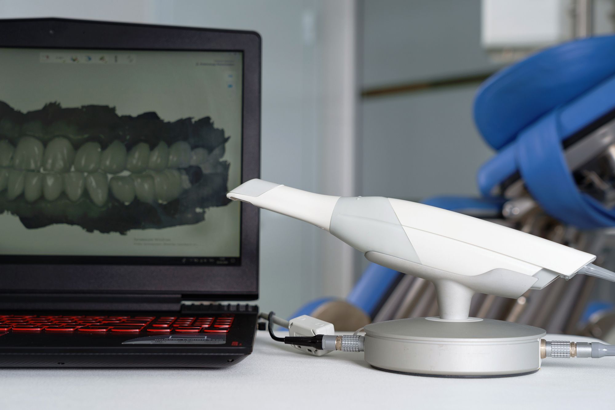
Understanding the Three Most Common Imaging Principles Used By Intraoral Scanners. Photo courtesy of Dental Pro Content/stock.adobe.com.
In many operatories, the intraoral scanner is the hardest working technology in the dental practice. The scanner can help patients see what you see, facilitate diagnosis ownership and treatment plan acceptance, and provide an excellent road map for restorative success with detailed and accurate digital impressions.
But do you know how it works?
Chances are you know how to use it, but not how it creates the images that do so many great things for your restorative cases.
Invented by Mörmann and Brandestini, scanners have been a part of patient care since 1985. However, the first scanners had problems with accuracy, particularly when scanning a wide area. The optical window of the scanner tip could only manage so much, so to complete the entire scanned area's image, the spatial data was aggregated, limiting the performance of scans to inlays or single crowns.1
However, advances in optical systems and imaging processes are changing, expanding scanners' capability. Scanners can be used for more extensive cases, like longer fixed dental prostheses and implants. They are also applicable to intraoral devices like surgical guides, individual trays for dentures, and metal frameworks of removable partial dentures.1
The 3 Technologies That Process Scan Data
Since 1985, more scanners have been on the market, competing for your business. No matter which brand you choose, a scanner has 3 parts that comprise its performance. These are the quality of the image capture, how it processes the data, and what it produces on screen. However, the critical element of performance is the imaging technology.2
The Scottish Dental Magazine says that an intraoral scanner's objective is to record the 3D Geometry of an object. This task relies on casting structured light upon an object from a handheld device, a sensor. The software then processes the images captured, creating a point cloud, which are the set of data points it collected about the object you are scanning. The software then analyzes the point cloud, creating a 3D surface model, aka a mesh, which the software outputs as an STL (which stands for stereolithography, or standard tessellation language or standard triangle language) file.3
Chances are, some of you knew that already. However, for the rest of us, it is important to note that are a few techniques that come together to make this happen. These include triangulation, confocal, and AWS ( active wavefront sampling). The Taiwanese Journal of Orthodontics describes them as:2
- Triangulation Technique: A scanner operates on the same idea as Trigonometry. The knowns determine the unknown, aka, the object's surface.3 The Pythagorean theorem (a2+b2=c2) plays a significant role in this area. The 3 points involved are the laser emitter, the sensor, and the object's surface. Since the software knows the distance and angulation of the images, it can determine the object surface.
- Confocal Laser Scanning: Point-and-Stitch reconstruction is a typical process to reconstruct the 3D image of the object you want to capture. The emitting laser filters through a tiny pinhole to the desired object. Then the scanner captures the light reflected from the object and only that object. Then using the 2D images from the different confocal planes, the software puts the image back into 3D (hence, the point-and-stitch part of the reconstruction).2
- Active Wavefront Sampling (AWS): This technique is the sampling method behind some scanners' 3D-in-motion video recording feature. Internal sensors (complementary metal-oxide-semiconductor, or CMOS) capture 3D information from different perspectives (triple imaging). The single-lens circles around a point of interest. The pattern produced by each point gives the software distance and depth information needed to make the 3D images.2
In addition, the Scottish Dental Journal says Stereophotogrammetry enhances the 3D imaging process. Stereophotogrammetry estimates the coordinates by analyzing two or more images, collecting data and information that defines the shape and location of the object in relation to other images from different angles. One way to think of this technique is the way you use binoculars. For example, the left side lens produces one image, the right another, which you can see when you shut the other eye. However, with both eyes open the images viewed together through the binoculars are one image.4 The technology uses an algorithm based on passive light projection and software. This approach is different from the active light projection and hardware involved in other parts of the process.3
Scanning: the Gateway to CAD/CAM
Among its many uses, the scanner kicks off the CAD/CAM dentistry workflow. After prepping and scanning, the 3D images created from the tooth preparation and surrounding teeth are how the dental team can design the final restoration before sending it off to the mill. The intraoral scan for a digital impression is better for the dental team's ability to deliver an efficient, well-fitting, and complete restorative process on the same day. In addition, it is a better patient experience to skip traditional impressions.
Alex Kalmanovich, DDS, ABDSM, a private practice dentist in Laguna Beach, CA, says that early in his career, he had the luxury of working in a practice that utilized new technology. One piece of their pie was a digital scanner and a milling machine. Dr Kalmanovich says the practice owner mentored him and taught Dr Kalmanovich how to utilize the technology successfully.
"When I opened my own practice, it made the decision just a little bit easier to make the big investment into digital dentistry. I knew I needed a way to stand out in the community, where multiple dentists are on every corner. So, although financially it was frightening, I had confidence that I could make a return on my investment," Dr Kalmanovich says.
Dr Kalmanovich chose the CEREC system by DENTSPLY Sirona for his private practice in Laguna Beach, CA. He describes CEREC as the Tesla of digital dentistry.
"Dentsply Sirona has relentlessly continued to push ahead, bringing the advent of digital dentistry into normal reality," Dr Kalmanovich says. "Whether you love or hate CEREC, they were instrumental in paving the way for many other companies to start producing digital scanners and milling machines. More practically, consider the availability of CAD/CAM dentistry in the early 2010s. Most dentists have considered buying a CEREC whether they admit it or not, leading to the world we live in now, where most dental practices have digital scanning equipment."
Dr Kalmanovich says it is essential that members of the dental profession be open to change. New technologies are going to continue to change the digital workflow. While it may seem daunting to incorporate new technology, especially when things they are doing now seem to work fine, Dr Kalmanovich encourages dental professionals to continue to adapt to it, enhancing the patient experience and improving treatment outcomes.
"As CAD/CAM and 3D printing are becoming common in many practices, I see in the future utilizing artificial technology and image recognition to help our treatment plan and monitor oral disease," Dr Kalmanovich says. "I love how dentists continue to be open to ways to improve our wonderful profession."
References
- Park, J., Shim, J., 2019, 'Optical Impression in Restorative Dentistry', in M. E.Machoy (ed.), Computer Vision in Dentistry, IntechOpen, London. 10.5772/intechopen.84605
- Hwang, Henry Hann-Min; Chou, Chi-Wei; Chen, Yi-Jane; and Yao, Chung-Chen Jane (2018) "An Overview of Digital Intraoral Scanners: Past, Present and Future- From an Orthodontic Perspective," Taiwanese Journal of Orthodontics: Vol. 30 : Iss. 3 , Article 3. doi:10.30036/TJO.201810_31(3).0003
- Patterson M. Intraoral scanners In dentistry – an update on digital technology - Scottish Dental magazine. www.sdmag.co.uk. Published 2018. Accessed September 20, 2022.https://www.sdmag.co.uk/2018/10/01/intraoral-scanners-in-dentistry-an-update-on-digital-technology/
- ŞAHAN N. What is Stereophotogrammetry? - Surveying Group. Surveyinggroup.com. Published 2020. Accessed September 20, 2022. https://surveyinggroup.com/what-is-stereophotogrammetry/


