• Best Practices New Normal
  • Digital Dentistry
  • Data Security
  • Implants
  • Catapult Education
  • COVID-19
  • Digital Imaging
  • Laser Dentistry
  • Restorative Dentistry
  • Cosmetic Dentistry
  • Periodontics
  • Oral Care
  • Evaluating Dental Materials
  • Cement and Adhesives
  • Equipment & Supplies
  • Ergonomics
  • Products
  • Dentures
  • Infection Control
  • Orthodontics
  • Technology
  • Techniques
  • Materials
  • Emerging Research
  • Pediatric Dentistry
  • Endodontics
  • Oral-Systemic Health

Technique Touch-Ups 2023

Publication
Article
Dental Products ReportDental Products Report June 2023
Volume 57
Issue 6

Experts from across the industry share their favorite tips, tricks, and solutions to make dental care better for patients and easier for you.

Technique Touch-Ups 2023

Technique Touch-Ups

May 25, 2023

Tip: Use CBCT to Educate Patients to Make Informed Treatment Decisions

From the most mundane decisions to the most important, we are used to having unlimited information at our fingertips to help decide whether we are making the right choice. So, when patients are told they need a crown or an extraction, they may balk. Ten years ago, there would have been no hesitation, but today Google, WebMD, and even Facebook are available for crowdsourcing information and opinions. It is more important than ever for doctors to use technology as a tool for educating patients about treatment while instilling confidence that the decision to proceed is right for them.

Ian Lowell, DDS - CBCT imaging in dentistry

Ian Lowell, DDS

Show, Do Not Just Tell

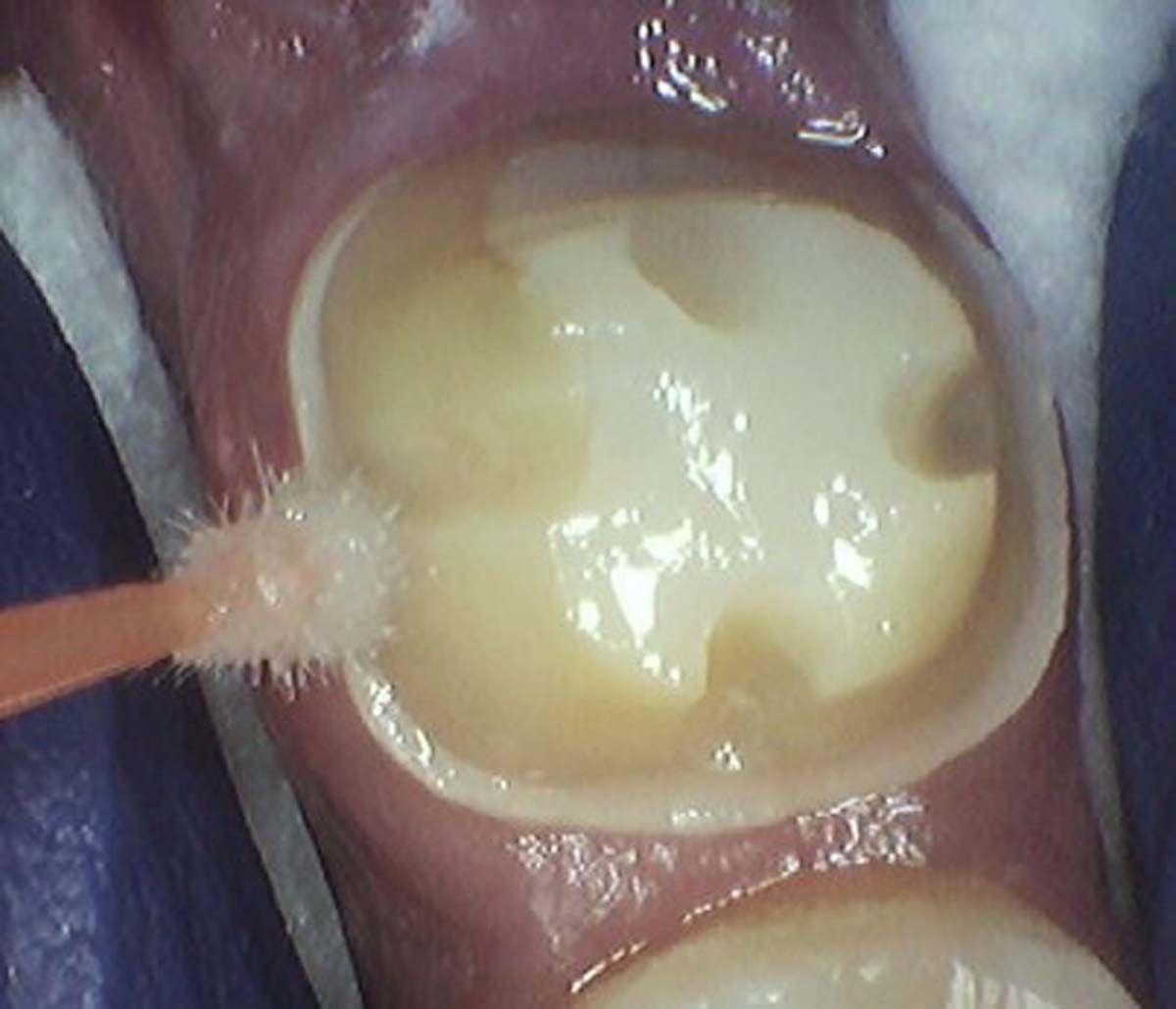
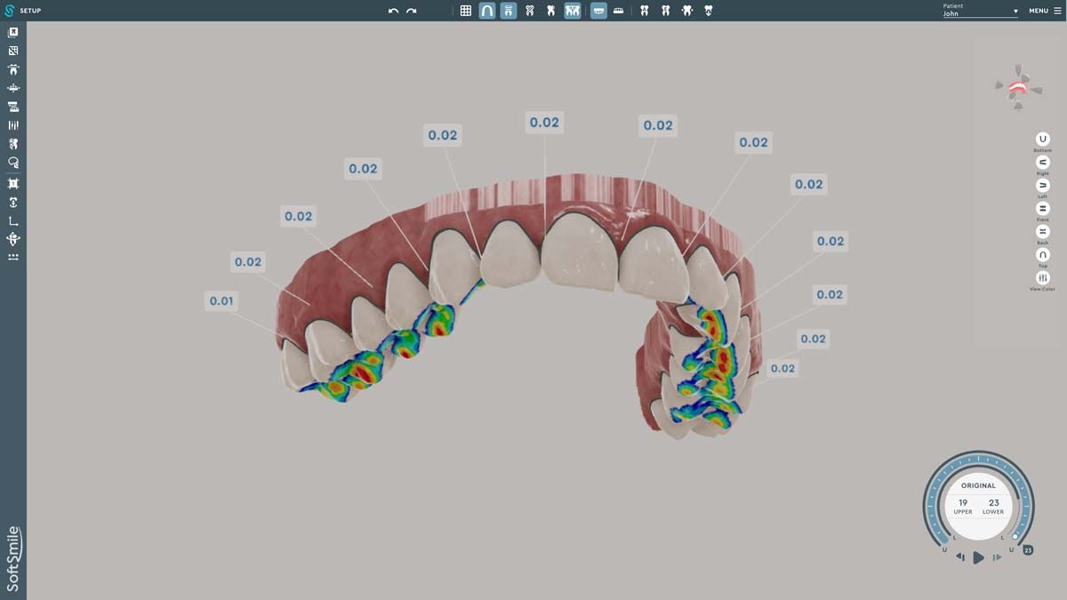
We used to try showing patients their clinical situation on 2D x-rays and demonstrating treatment using models. Later, digital intraoral cameras helped, but they could not show much more than what a patient could see in a mirror. Today, it is possible to show patients every angle of their clinical situation in hyperrealistic 3D with a CBCT scan. Now, when I recommend treatment to patients and they ask why, I am armed with an interactive image captured by my CS 8200 3D extraoral imaging system (Carestream Dental). I scroll through the tooth and bone, point out lesions below the surface, and show why there is or is not enough bone for an implant. If you use something like the Prosthetic-Driven Implant Planning module to automatically merge an intraoral scanner file with the CBCT scan, you create an even more accurate picture. How much easier is it for the patient to accept treatment when they are seeing the end results of that treatment?

CBCT imaging in dentisry

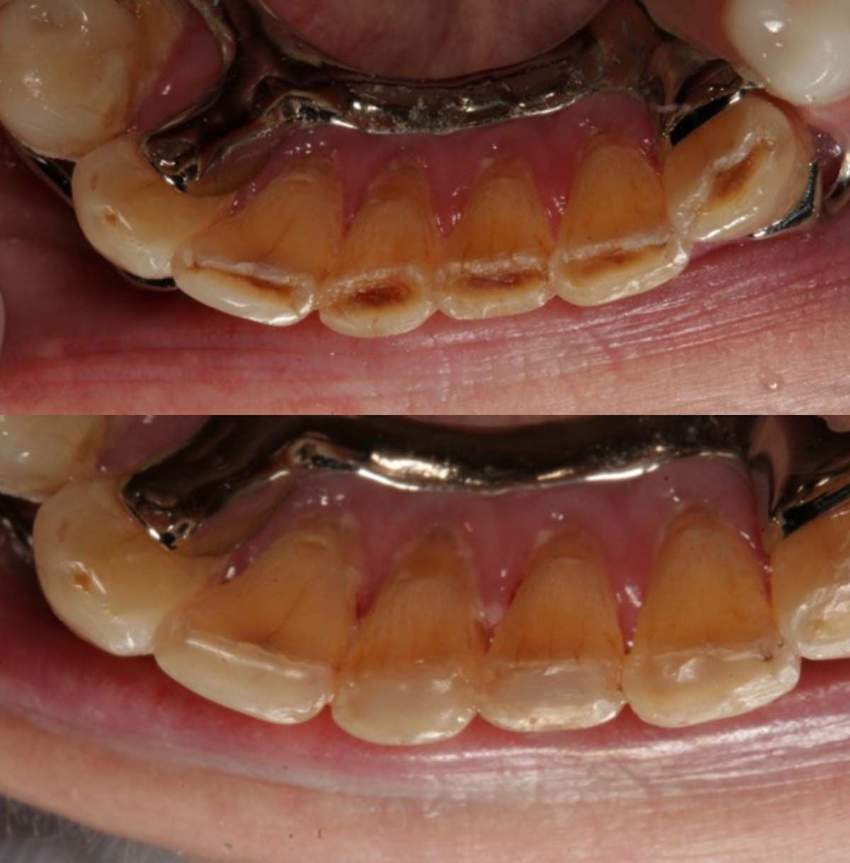
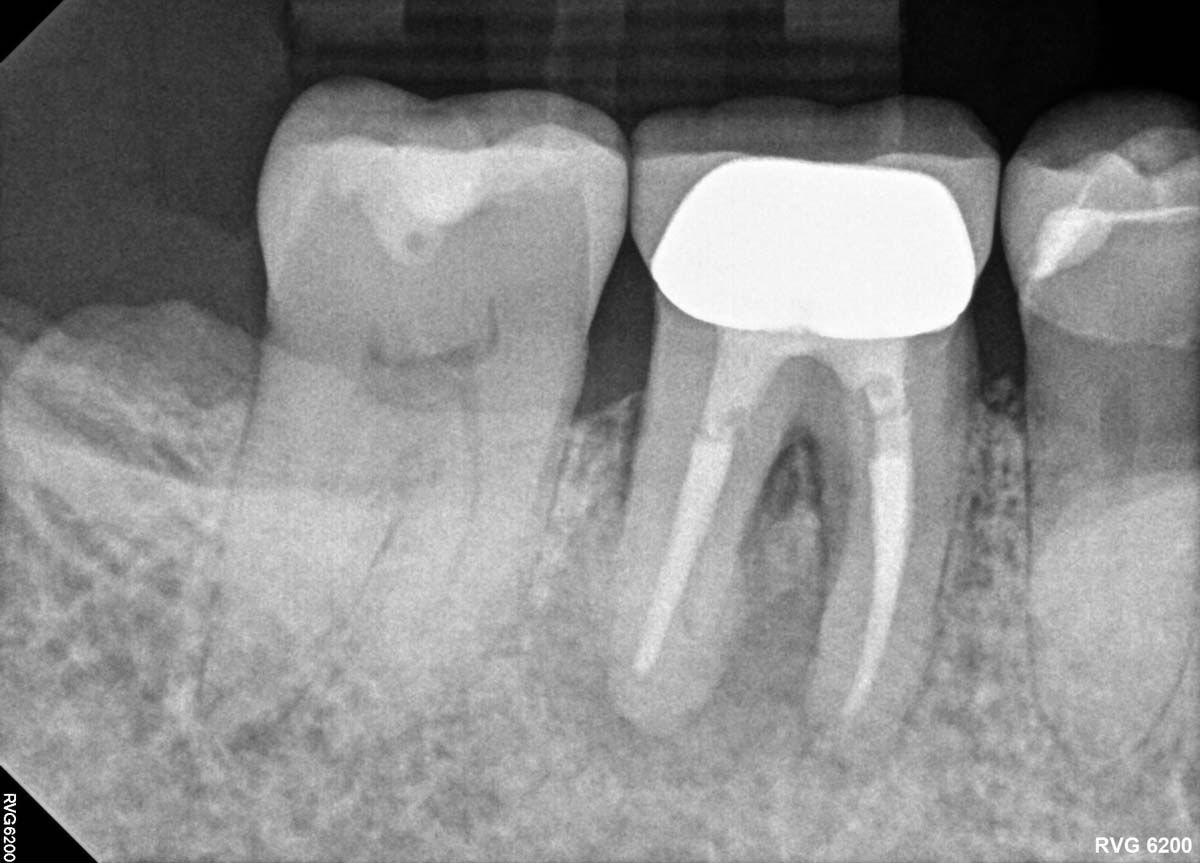
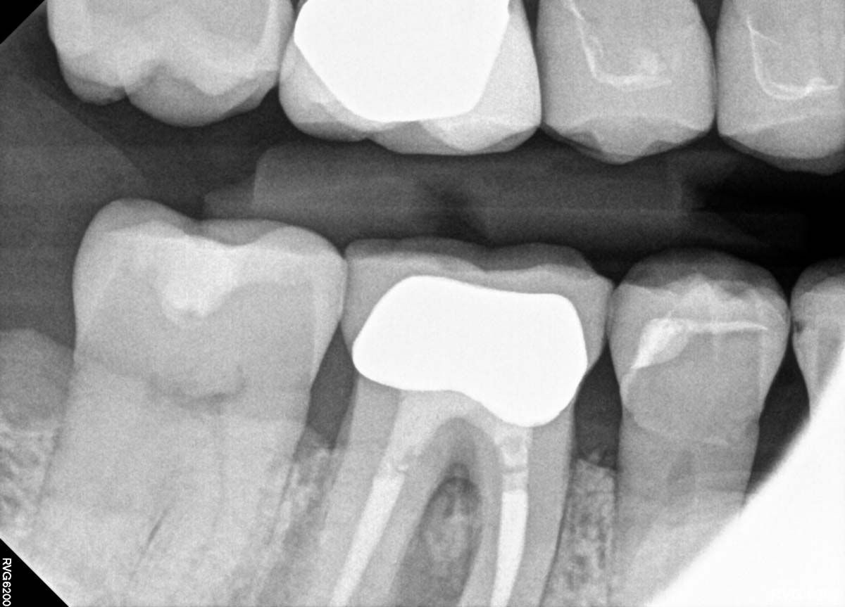
Infection was identified on pariapical x-rays.

Prepare Your Patients

Suppose CBCT has revealed that the issue your patient is experiencing requires a specialist. A 3D scan tells me when it is best to refer treatment, but that can be frustrating and confusing to a patient who was not expecting a second appointment at another office. In that case, the 3D scan lets me prepare patients for the next steps. Yes, they are being sent to a specialist, but they will have realistic expectations about what the next doctor might find and recommend for treatment. Because I also send the scan to the specialist, there are no surprises for anybody; patients do not feel like they are arriving at another office only to be blindsided.

A look at patient's periapical x-rays.

A look at patient's periapical x-rays.

Give Peace of Mind

Oral health care is a negative-demand business. We are telling patients things they do not want to hear. Time, money, and often patient discomfort are involved. You never want a patient leaving your practice wondering what they just agreed to—or, even worse, declining the right treatment because a 2D panoramic image did not look “that bad.” When you use a CBCT system to diagnose, plan treatment, and inform patients about their care, they do not only have confidence in their decision to accept treatment; they have more confidence in you.

Use CBCT  to Educate Patients to Make Informed Treatment Decisions

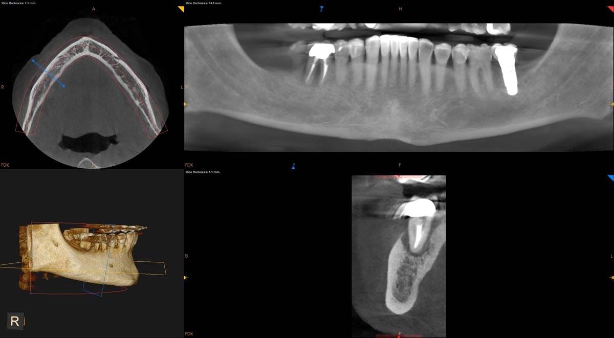
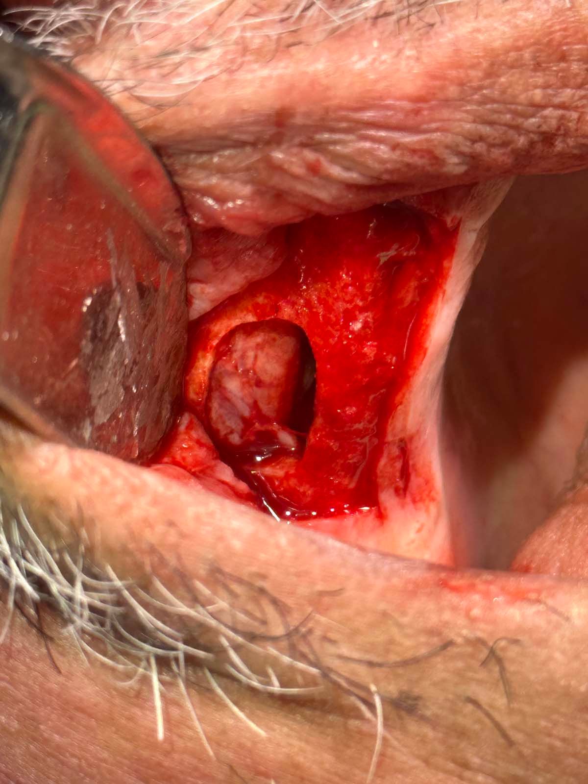
The CBCT revealed the extent and solidified the opportunity to make the correct clinical decision

Informed consent is more than a patient signing a document, and it is our job as practitioners to provide patients with as much diagnostic information as possible to make that informed decision. When patients feel like they have all the information needed to understand why I am recommending a certain path forward, I know I have done my job.


May 25, 2023

Tip: Choose a Reliable and Esthetic Composite for Clear Aligner Therapy

Clinical photo by Dr. Stephanie Vondrak Vondrak Dental  Elkhorn, NE

Photo courtesy of Dr Stephanie Vondrak, Vondrak Dental, Elkhorn, NE.

In 1998, clear aligner therapy was approved by the United States Food and Drug Administration, expanding the scope of services for general dental practices. This was a new, easily accessible form of orthodontics that general dentists could implement without the inventory or education required by traditional brackets and wires. To date, it is estimated that more than 14 million smiles have been treated with clear aligner therapy.

As a health-centered dentist, I routinely offer clear aligners to eliminate crowding (plaque traps), level occlusal planes, stabilize occlusion, and/or correct rotations prior to complex cosmetic or rehabilitative treatment plans. Clear aligners are a fantastic tool. However, clear aligner therapy is not without challenges.

Clear aligner therapy requires staff education, scanning equipment, and large quantities of PVS material as well as patient adherence for optimal results. For me, however, the most significant challenge has been mastering the technique for composite button or attachment placement. Without a consistent system for attachment placement or a reliable composite material like OMNICHROMA (Tokuyama Dental America), this step of clear aligner therapy is lengthy, frustrating, and expensive. Likewise, composite buttons can be a “deal-breaker” with treatment acceptance if attachments are to be placed in the esthetic zone (the upper front 6 teeth). Following are some helpful tips, especially in easing esthetic concerns around treatment acceptance with the “picky patient.”

Most know that when placing attachments, isolation is essential. A dry field must be established to facilitate the best bond to your patient’s clean, pumiced enamel surface. Techniques like placing a flexible, latex-free lip retractor prior to pumice, etch and bond are well known. The most significant tip I can offer is in choosing the most reliable and esthetic composite system: OMNICHROMA.

OMNICHROMA composites are based on structural color and eliminate the need for shade matching, reflecting the shade of the underlying enamel surface. They are offered in several viscosities, so you can choose one of the OMNICHROMA products that works best for you: packable OMNICHROMA, OMNICHROMA Flow, or OMNICHROMA Flow BULK. I study the clear aligner template; prior to placement, I look at the shape of the attachment to be placed and choose accordingly. If the attachment is large or I am concerned it might have a sharp edge, OMNICHROMA Flow and OMNICHROMA Flow BULK work well. When cured, both are smooth and require little polish. This is especially helpful, for example, when placing an attachment on the facial surface of an upper second molar or rotated premolar. Likewise, I prefer packable OMNICHROMA for placing smaller attachments with a rounded edge. These attachments are often seen on anterior teeth. The milky white appearance of OMNICHROMA products when placed disappears when cured, and the structural color of OMNICHROMA, no matter the viscosity, seamlessly blends onto the enamel surface every time.

Thanks to a reliable and esthetic composite system like OMNICHROMA, the “picky patient” is happy with the clear aligner results and the process. In addition to the esthetic results you can achieve with OMNICHROMA, you can avoid the expense of purchasing multiple shades of composite as well as wasting chair time with the shade-matching process.

[In practice for 20 years, Dr Vondrak owns 3 dental practices in the Elkhorn, Nebraska, area. Advanced services offered in her practice include temporomandibular joint therapy, therapeutic Botox injections, sleep apnea appliances, and more.]


May 25, 2023

Tip: A Simple Plug-and-Play System Can Make Your Favorite X-ray Sensor Wireless

Air2Zed is a simple plug-and-play system that makes your favorite x-ray sensor wireless

Becoming a dentist is a great accomplishment, but this is not the end of the road; in fact, it is just the start. Since I was young, it was always an innate process on how I could make something more efficient—even something simple like using my time efficiently from the second I woke up to when I left for school. I have always tried to utilize the maximum potential in any given situation so I can have more time for extracurricular activities.

This characteristic is equivalently important in dentistry. Running an efficient practice through techniques and technology has allowed me to work 3½ days a week. You must work harder to make systems simple, but ultimately the results speak for themselves. I have invested in both my employees and technology to create a simple yet efficient workflow in the office.

As Benjamin Franklin famously said, “An investment in education always pays the highest returns.” I invest a lot of hours in educating my employees on processes and procedures in dentistry and technology so they can help the patients, the staff, me, and the office. We have set up simple, efficient, and replicable systems for each employee to run the office seamlessly. One of these is the Air2Zed wireless x-ray system.

Air2Zed is a simple plug-and-play system that makes your favorite x-ray sensor wireless. The battery on the system lasts all day and there is no additional software to install. Within 2 minutes or less, your staff will know how to use this system, so there is no need to block your productive work schedule for hours or multiple days of training.

Air2Zed has eliminated the clutter of having extra wires while preventing accidental damage to sensors from dropping them or tripping over wires. A dentist friend of mine recently had 2 broken sensors in the same day. Imagine the loss of revenue this can cause. This easily could have been prevented if they had incorporated a wireless imaging workflow.

From start to finish, it takes my assistant 5 minutes to take an FMX on a patient. There is an immense return on investment with this technology. My assistants are happier to take FMX, the shorter time taking x-rays allows more patients to be seen, and the sensors have not been dropped or tripped over since Air2Zed was welcomed in our office.

Bill Gates said, “The first rule of any technology used in a business is that automation applied to an efficient operation will magnify the efficiency. The second is that automation applied to an inefficient operation will magnify the inefficiency.” Creating an efficient system that is also simple requires quite a bit of thought and effort. Air2Zed has created a technology that incorporates this principle, making it simpler for clinicians to apply it in the office while making their smart investment even smarter.


May 25, 2023

Tip: Hidden Income From Patients Already in Your Chair

The before and after clinical photos courtesy of Tony Tomaro, DDS,Chicago, Illinois.

The before and after clinical photos courtesy of Tony Tomaro, DDS,Chicago, Illinois.

The demographics of the dental patient have changed. Patients are living longer. With lifetime expectancy increasing, we see many more patients with worn dentition, perhaps the result of clenching, grinding, or erosion over the years. Predominantly seen in the aging population, this is also observed in younger patients, which is concerning if not diagnosed early. The clinical manifestation of wear or erosion can result in decreased vertical dimension; flattening of the incisal edges; facial, cervical, incisal, and occlusal abfractions; chipping of teeth throughout; worn enamel resulting in exposed dentin; sensitivity; and perhaps more.

“Aging is not lost youth but a new stage of opportunity and strength,” writer Betty Friedan said.

One of those areas of opportunity would be the treatment of anterior incisor abfractions and worn incisal edges. Mandibular incisors may show chipping and breakdown of the lingual enamel, smooth buccal enamel, and remnants of the pulp that may have receded or calcified. Stains and decay are common.

The key, as in any treatment acceptance, would be to present the benefits of such treatment, as follows:reduce and slow breakdown and wear of teeth;strengthening teeth when the abfractions are restored;minimally invasive;benefits of a bioactive filling material; reduce sensitivity;improve esthetics; and in majority of cases, no anesthetic is required.

Preparation Technique

The goal of anesthetic-free minimal preparation can vary slightly between decay or stain present. For teeth with no decay or stain present, bevel the incisal edge and remove any irregular enamel chipping with a fine or extra-fine finishing flame diamond bur with reduced speed of the handpiece. Roughen the dentin of the abfraction with a small carbide bur, such as a 330 or 556 fissure bur. If decay is present and no anesthetic is used, cautiously remove the decay with a brush technique. A reduced speed in the handpiece using the same burs will reduce the possibility of discomfort or pain in the patient. If there is staining and no decay, the clinician will determine whether stain should be left, removed, or partially removed. To date, I have not had any patient mention pain during this procedure. Upon completion of the preparation, I will cleanse the preparation with 2% chlorhexidine.

Restorative Materials

My preferred material is a flowable composite for these conservative procedures. With the availability of many flowable composites, I want one that adapts well and bonds to the preparation, is easy to place, maintains strength, is easy to polish, and has bioactive properties. Mostly, bioactive materials are either calcium silicates or calcium aluminates, both of which are set with an acid-based chemical reaction to produce an alkaline pH. Shofu provides the clinician with ideal flowable composites with all these characteristics and more with Beautifil Flow Plus (value priced) and Beautifil Flow Plus X (premium). These nanohybrid flowable composites come in 2 viscosities (low and no flow) and multiple shades, are durable, and contain GIOMER Technology.

GIOMER Technology is bioactive technology that releases and recharges 6 beneficial ions, including fluoride. Together, these ions have been clinically proven to inhibit plaque, neutralize acid, and eliminate secondary decay.1 They reduce tooth mineral solubility, encouraging the remineralization of tooth enamel and decreasing the acid production of cariogenic bacteria.2

After completing the preparation and proper isolation, Shofu provides the clinician with a self-etch, 1-component bonding agent, BeautiBond. This unique bonding agent allows the clinician different etching techniques: total, selective, or self-etch. My preference is selective etch. The enamel is etched with 35% phosphoric acid for 15 seconds, rinsed, and dried. BeautiBond is applied and left undisturbed for 10 seconds. Dry with a gentle stream of air for about 3 seconds and then dry with stronger air until a thin, uniform bonding layer is obtained. Light cure with an LED light curing unit for 5 seconds. Using Beautifil Flow Plus X with enhanced polishability, I apply the low flow viscosity (F03) that provides self-leveling and adaptability. Check the occlusion and adjust if needed. The final restoration was finished with Shofu’s Super-Snap X-Treme Polishers and CompoSite Polishing Paste. With the application of Beautifil Flow Plus X, we have a final restoration with high flexural strength, low wear resistance, low shrinkage, and tight marginal seal. Nanohybrid particles provide a high level of polishability and a chameleon effect, esthetically blending with natural teeth, that dentists have come to expect.

Using an anesthetic-free procedure, reducing wear, and improving and/or maintaining strength of the teeth with an esthetic result will make the dentist and dental team heroes to the patients.

References
1. Gordan VV, Mondragon E, Watson RE, Garvan C, Mjör IA. A clinical evaluation of a self-etching primer and a giomer restorative material: results at eight years. J Am Dent Assoc. 2007;138(5):621-627. doi:10.14219/jada.archive.2007.0233
2. Ozer F, Irmak O, Yakymiv O, et al. Three-year clinical performance of two giomer restorative materials in restorations. Oper Dent. 2021;46(1):E60-E67. doi:10.2341/17-353-C
3. Rusnac ME, Prodan D, Cuc S, et al. Water sorption and solubility of flowable giomers. Materials (Basel). 2021;14(9):2399. doi:10.3390/ma14092399
4. Giacaman RA, Muñoz-Sandoval C, Neuhaus KW, Fontana M, Chalas R. Evidence-based strategies for the minimally invasive treatment of carious lesions: review of the literature. Adv Clin Exp Med. 2018;27(7):1009-1016. doi:10.17219/acem/77022


May 25, 2023

Tip: AI Technology Is Making an Impact on Diagnosis, Patient Communication

VideaHealth’s AI program for caries detection,

I often joke with my patients about how frustrating dentists can be. I explain that if you show 10 dentists the same information, you may get 10 different opinions on treatment recommendations or lack thereof. The diagnostic information we gather feeds our treatment planning, but interpreting those diagnostics leads to sometimes dramatic differences in opinion. Especially in a group practice setting like mine, how do we embrace different dentists’ creativity, experience, and perspective while keeping consistent foundational diagnostic data for our patients? Taking it further, how do we convey this information to our patients with varying dental knowledge and insurance companies looking for reasons to say no to treatment we feel certain is right and necessary for patients?

Nearing the end of 2022, our practice had the opportunity to pilot VideaHealth’s AI program for caries detection, which now includes capabilities for radiographic bone loss and periapical lesions. What I find exciting about the product is it gives multiple clinicians the same set of data that we can then layer into our own clinical experience—or, in the case of a new dentist with little experience, immediate data to aid further assessments. With caries or periapical pathology, red boxes denote areas for evaluation. For radiographic bone loss, varying degrees of disease are noted with green, yellow, or red to indicate severity, with associated measurements listed. We can discuss as professionals what treatment to recommend with these data, but the program standardizes information, which has been challenging in dental clinical assessment.

Now we have standardized data that opens a conversation for dentists to layer in experience and clinical preferences, but the program also creates an important visual tool for our patients. It quickly notes areas for further discussion. It saves time for noting areas of concern and having to draw or point to nuances on our imaging. If we do not agree with providing treatment for all highlighted areas, it offers an opportunity to explain disease progression to patients. For example, suppose it notes an area of decay that is still in enamel that we do not yet advise restoring. In that case, it can strengthen our patients’ confidence in our assessment as we exhibit our ability to utilize technology while balancing other clinical knowledge and considerations. It is hard to argue that red, yellow, and green markings on the radiographs are not an impactful tool for discussing a disease process that can be very confusing for patients.

Lastly, we need to consider how this can affect treatment communication with dental insurance companies. The process of communicating for treatment preauthorization or payment is a tremendous drain on our office. The time and expense that go into trying to collect on procedures that practitioners feel confident our patients need is a resource drain—not to mention that when dental insurance companies deny treatment coverage, patients often look to us as the “bad guys” and infer treatment was unnecessary despite how obvious we find the procedure indications. VideaAI allows for an automated narrative to be written and attached to claims with clearly marked, noted, and measured information.

It is an exciting, cutting-edge tool that has been an important clinical aid for our doctors in disease process detection and helpful for communication with insurance companies and patients.


May 25, 2023

Tip: Switching to Digital Can Deliver Beautiful, Strong Dentures

The PrograMill PM7 from Ivoclar set up at Hatch Dental Lab.

Luckily, the terrible old days of making analog-style dentures are in my past. That involved shrinkage, dentures breaking, porosity, training staff, and too many more problems to list. I will never go back.

In 2019, we partnered with Ivoclar and transitioned our lab to digital. The Ivotion Denture System has improved my life. I have evenings and weekends back with my family, now that I have digital equipment doing most of the manual labor. The dentures are beautiful and extremely strong. Any lab or office wanting to make digital dentures should look at many of the problems they are having with analog, and most of them will likely be solved.

A main concern I hear from offices that start working with us is, “I like my chairside workflow and I don’t want to switch it up.” The beauty of digital dentures is that chairside workflow can stay the same. Many individuals talk about getting dentures in 2 appointments. That is possible, but do you get the best outcome that way? We do not. We see the best outcomes when offices do initial impressions, final impressions, vertical dimension of occlusion (VDO), and a try-in appointment before delivery. The principles of making a denture are the same, and this is important to remember. The beauty of digital is that lab errors are minimal.

What makes a digital denture succeed and what makes one fail? The chairside record appointment needs to be perfect. If the bite is off, the vertical is too open or too closed, the midline is off, or the high lip line or incisal edge is off at the VDO, then these also will be off at the try-in. Digital will precisely replicate the record you take chairside. We hear frequently from dentists, who have accurate records, that they have never seen better occlusion or a better fit.

Dentures milled with the PrograMill PM7.

Dentures milled with the PrograMill PM7.

We have some of our offices do a single try-in more than 90% of the time, and we have offices that do multiple try-ins more than 90% of the time. What makes the difference? When I talk to the offices, they usually say, “My new assistant is doing the VDO,” or, “The patient was anxious to get out of the chair, so we had to hurry.” Again, if the VDO is not accurate, there is not a magical digital button to fix this in the lab. This is where the dentist’s expertise is important. Because we are following the wax rims and bite registration, there is no indication to the lab if anything is off. It is much more frustrating to patients and offices to have a patient come for a try-in when the fit is not even close. Patients quickly lose confidence, and it costs the office and patient time and money.

Treat the VDO like a try-in appointment. Have the patient look at the wax to ensure they like the extent to which teeth will be showing, show them the midline, and check multiple times for a solid, accurate bite. If the patient is not consistently closing into the bite you just took, spend a few minutes determining why. Taking an extra 10 minutes at the VDO saves everyone time.

Digital dentures, and specifically the Ivotion Denture System by Ivoclar, have changed my life. They are extremely accurate and esthetic. Your office and patients will love them as well. I would quit the dental business before ever going back to analog


May 25, 2023

Tip: Choose a Family of Composites That Can Bring You Highly Esthetic Restorations

All 3 CLEARFIL MAJESTY systems from Kuraray offer a chameleon effect and LD technology.

All 3 CLEARFIL MAJESTY systems offer a chameleon effect and LD technology.

Mirror, mirror, on the wall: Who has an esthetic composite with choices for all?

There is a reason Kuraray Noritake calls its composite “Majesty”—it is formulated to reign supreme and deliver dignity and beauty. So how do you pick? Let us try to simplify it for you.

CLEARFIL MAJESTY ES-2 Universal:

This material is 78wt% filled and is available in syringe and PLT (preloaded tip) delivery systems. Because it is available in only 1 shade, the best application is in the posterior region of the mouth. A chameleon effect and LD (light-diffusion) technology make for seamless blending and almost indetectable margins. Inventory can be streamlined, because only 1 shade choice must be made.

CLEARFIL MAJESTY ES-2 Classic:

This material is also 78wt% filled; it is available in the same delivery systems as CLEARFIL MAJESTY ES-2 Universal. There are 16 traditional shades available, ranging from A to D, with the addition of 2 bleaching shades: XW and W. The material is intended to be used as a monolayer product, selecting 1 specific shade for the restoration. The science in shade selection is based on lightness, utilizing the VITA Classical Shade Guide. We recommend removing all the shade tabs from the guide and arranging them from lightest to darkest. This will help when conflict arises between 2 shade choices. You can view the shade to the right or the left of the center tab (always an A) to determine the most accurate shade match.

CLEARFIL MAJESTY ES-2 Premium:

This offers the same characteristics and delivery systems as mentioned previously. The major difference is that this is a multilayer product; a selection of both an E and a D shade will be used on each restoration. Once the guide is rearranged from the lightest to the darkest shade, you will notice that every group of 3 tabs has an A tab in the center. When using Premium, you will see that only 5 A choices are available for ordering, with a corresponding E and D offered. As an example, if you select shade A2, you would use A2E on the enamel portion of the tooth and A2D on the dentin portion. This product offers a more esthetic effect and is loved by biomimetic dentists.

All 3 systems offer a chameleon effect and LD Technology. The choice is yours—each can bring highly esthetic restorations your patients will love, so you can treat your patients like royalty.


May 25, 2023

Tip: Educate Your Patients Through Images and Teledentistry

Integrating an affordable intraoral camera, such as MouthWatch, into every operatory of clinical practice is easy and benefits the dental provider and patients.

The first and most effective step to case acceptance and changing patient behaviors is through education. Allowing your patients to see what you see through the lens of an intraoral camera enhances their education and understanding. As a result, they often become more invested in their oral health journey. Although intraoral cameras have been around for decades, they are now affordable for each operatory and patients often perceive them as innovative technology. A small piece of equipment can have a big impact on your patients and practice.

Integrating an affordable intraoral camera, such as MouthWatch, into every operatory of clinical practice is easy and benefits the dental provider and patients. With practice, a full series of intraoral photographic images can be captured in approximately 90 seconds and incorporated into all parts of the patient experience, which may lead to improved health outcomes. Simple tricks include drying to the tooth or quadrant on which you wish to capture images and working around strategically, like the sequence you use to capture radiographs. Ensure the dental team has time to get familiar with any piece of equipment prior to patient use to avoid uncertainty.

Improving the efficiency of the dental team when discussing oral health risks and outcomes, presenting treatment plans, and submitting claims can be achieved with the use of intraoral imaging. When discussing with patients, sit them upright and show the intraoral image side by side with a radiograph to demonstrate the oral condition and treatment recommendations. A full series of intraoral images should be obtained on all new patients to establish a baseline and can be used for comparison, referrals, and claims support.

Intraoral imaging is for improved education, increased efficiency, and teledentistry. The dental experience is evolving, driven by patient demand for convenient telehealth options, and this includes oral health care. The ability to provide a convenient, effective dental visit meeting patients’ needs wherever they are sets the stage for happy patients. Dental offices also reap the reward of teledentistry when increasing efficiency by completing virtual visits, such as postoperative and limited evaluations.

Although cameras and teledentistry are not tied together, each complements the other in achieving patient care. Intraoral cameras in combination with teledentistry open the possibility of expanded hygiene hours, on-demand referrals and consultations, and alternative practice models. You can streamline efficiency when the average teledentistry visit is 15 to 20 minutes vs a 30-minute in-office limited evaluation. By offering a virtual option, you eliminate the need for travel to an in-office exam only to return for treatment later. Teledentistry enables dental practices to determine patient needs and schedule accordingly, reserving chair time for production and potentially serving as a practice builder.

Intraoral camera imaging is considered a mainstream and expected part of a dental appointment by many patients. They are more invested than ever and expect a high level of service and information from their health care providers. Incorporating intraoral imaging and teledentistry can be a convenient, effective service that sets your practice apart.

[Jamie Collins, BS, RDH-EA, won the American Dental Hygienists’ Association (ADHA) Trailblazer Award in 2021 and 2022 and was the first recipient of the ADHA Standout Seven Corporate Hygienist Award in 2022. She is the sales and education manager at MouthWatch and can be contacted at jamie@mouthwatch.com.]


May 25, 2023

Tip: Use Change Management, Latest Technology to Ensure Optimal Outcomes

The TRIOS intra oral scanner from 3Shape.

Throughout my time as a dental professional, I have been fully committed to ensuring that each clinical patient outcome is excellent. High-quality patient care is at the center of my ethos, and I find great joy in being part of a team that ensures that Aspen’s more than 1000 locations across the United States are delivering it. Staying up-to-date with technology is important; new devices can help you create better dentures while delivering optimal care.

3Shape has developed award-winning dental lab and intraoral scanners. These allow you to scan physical impressions within a patient’s complete removable prosthesis effectively and efficiently. Combining this innovative technology with the reference denture technique, which is preferred by clinicians and technicians for its clinically integrated workflow, gives dental professionals a comprehensive scan strategy to follow. This strategy delivers increased patient acceptance and satisfaction, reduces postoperative adjustments, and limits the need for multiple visits. With fewer clinic visits required for denture patients, you can free up chair time and provide care to more patients. At Aspen practices today, we have a high success rate at our denture delivery appointments. If we are providing 100 dentures per month, we have freed up 80 appointments, which translates into reduced patient wait times and better dentistry.

The digitalization of the denture process also allows electronic preservation of a patient’s scans and digital denture design. This means that if patients lose their dentures, they can have a new, identical set manufactured for delivery without having to visit the clinic.

With the change in technology comes a shift in denture manufacturing material. For decades, the standard denture material has been polymethyl methacrylate (PMMA), which has provided removable therapy to millions of patients. Today the industry is moving toward additive manufacturing, otherwise known as 3D printing, which uses a material with a different chemistry than PMMA. The combination of new technology and new materials can produce dentures and other removable prosthodontics that are stronger and more consistent, more easily customized, and that fit patients’ mouths as naturally as possible.

Although there is a cost associated with transitioning to digital denture technology, my advice for dentists is to find a lab or manufacturing center using this technology and partner with them on a few cases. You will be able to reduce your risk as you get comfortable with the technology, and then you can make the investment. Seeing really is believing.

My final tip for moving toward a digital ecosystem for dentures and other removable prosthodontics therapy would be to focus on change management. Often, new technologies and techniques face resistance because of inadequate introduction and training. Change is hard, especially when we think what we are currently doing works fine. Creating educational materials, whether they be hands-on training programs or e-learning modules, can help dental professionals get on board with this new technology. We want to provide the best care and experience for our patients. Any innovative technology or techniques that let us do that deserve our attention and consideration.


May 25, 2023

Tip: Select a Financing Tool That Helps You and Your Patients

Sunbit can offer more patients financing for their dental treatment by using artificial intelligence and machine learning technology.

Many dental practices today face common pain points, but 2 preventable issues have emerged as particularly troublesome. One is the difficulty in hiring, training, and retaining front office staff due to the labor shortage. The other is the inconsistency in presenting treatment plans, which leads to fewer completed plans and to subpar patient experiences.

To address these issues, I recommend creating a consistent, repeatable, and simple operations playbook that outlines processes, policies, and standard operating procedures. Eliminating inconsistency is crucial, as it leads to higher cost of running a business, lower revenue, turnover, burnout, and poor experiences for your team and patients.

A playbook does not have to be a monumental effort. By creating something useful and clear in a short time, you can address these problem areas and expect positive outcomes. Everyone in the front office quickly understands practice priorities, offering consistent experiences regardless of tenure. Bias is eliminated, ensuring every patient receives the same white-glove experience when presented with treatment plans and payment options. Financing is offered to every patient, and not just based on perceived need.

Our practices use Sunbit as the financing tool of choice. Our team knows that virtually every patient can get approved with fair rates and no fees. The process is fast, easy, and puts customers first, aligning with our high standards for patient experience. Sunbit also provides support to our team, with online training courses for new hires and ongoing training and engagement opportunities for existing employees via newsletters, webinars, and contests.

From the moment someone walks into the practice until they leave, your front office team has a significant impact on their experience. A thoughtful, well-designed, and simple playbook gives your team the chance to be successful, reliable, and fair, contributing to the bottom line and making everyone happy. Consistency is the key to success, and eliminating inconsistency will benefit your team and patients.


May 25, 2023

Tip: Revolutionize Dental Implant Practice With the W&H Piezomed Module

Tip:Revolutionize Dental Implant  Practice With the  W&H Piezomed Module

In the ever-evolving world of dentistry, professionals are continuously seeking innovative ways to improve clinical outcomes, increase workflow efficiency, and provide the best possible care for patients. Among the pioneers of modern dental implant practices is Joe Mehranfar, DMD, MS, a practicing dental professional and clinical director of the Implant Education Company, LLC, in Phoenix, Arizona. One such game-changing tool that Dr Mehranfar has utilized is the Piezomed module (W&H).

The Piezomed module has significantly improved workflow for dental implant procedures, allowing surgery to be completed sooner while achieving excellent outcomes. The device features a compact footprint providing ample room for my other instruments on the table, which is essential given limited operatory space. By integrating the Piezomed module with the Implantmed system, I can easily switch between rotary and piezo during the surgical procedure, resulting in a seamless, efficient workflow. This allows me to provide the optimal patient outcome and to remain focused on each patient’s specific needs instead of which equipment the team did or did not bring into the operatory.

The Piezomed module helps me to deliver excellent clinical care and simplifies the overall process for my team. Gone are the days when it was necessary to bring a separate piezo surgical device out of the closet for specific procedures. With the Piezomed module, I can keep my focus on the patient, resulting in a more efficient, less stressful experience for everyone.

In addition to the small footprint and simplified operatory setup, the Piezomed module is cost-effective. The system utilizes 1 saline bag and 1 irrigation line, eliminating the need for 2 irrigation lines (from 2 suppliers), lowering the overall cost for each procedure. Furthermore, because I am already using W&H surgical handpieces on every patient, the Piezomed module’s compatibility with the surgical unit adds another layer of convenience. The Piezomed module and the surgical handpieces are controlled with a single foot pedal, streamlining each case.

The Piezomed module in use in the author’s practice when performing a dental implant surgery.

The Piezomed module in use in the author’s practice when performing a dental implant surgery.

The Piezomed module boasts an innovative plug-and-play system that automatically detects each tip that is torqued onto the handpiece, setting the appropriate power and irrigation levels, adapting to the requirements of each procedure. This advanced technology eliminates the need for manual adjustments commonly associated with other devices, ensuring a smooth, efficient process from start to finish.

The Piezomed module is revolutionizing the field of dental implant procedures. Its integration with the Implantmed system, compact design, cost-effectiveness, and innovative plug-and-play technology have significantly improved the workflow, supporting my ability to provide exceptional quality of care to patients. By simplifying the process without sacrificing performance, the Piezomed module is proving to be an invaluable tool in my practice when performing dental implant surgery.

[Dr Joe Mehranfar is clinical director of the Implant Education Company, LLC, a full-service training center in Phoenix, Arizona. He is also clinical director of Dental Hearts, a nonprofit dental implant center, and co-owner of Aria Dental Implant Center, both in Phoenix.]


May 25, 2023

Tip: Digital Impressions Tips for Digital Dentistry Workflows

Eric Mayuga, DDS, uses the Roland DGA DGSHAPE DWX-42W chairside milling solution.

Eric Mayuga, DDS, uses the Roland DGA DGSHAPE DWX-42W chairside milling solution.

Digital impression capability is a must-have for every dental practice. It is a wonderful tool for patient education, comfort and convenience, and productivity for the dental team.

If you already use an intraoral camera, then you will quickly learn to use a scanner for digital imaging and impressions. At the first visit, we take a full-mouth radiographic survey and multiple intraoral pictures with our cameras. We have recently started to take digital impressions as a standard on adult patients and find that having them at the first visit is beneficial for the patient as well as for our office. Intraoral pictures are the standard and patients can see what we see. However intraoral pictures are static. You cannot manipulate the pictures other than changing colors and contrast. With a digital impression, we can now show a patient their dentition in 3D. We can manipulate the arches at any angle we choose and can show the patient how their teeth interact. This is important to show, for instance, malocclusions and detrimental effects of bruxism. When indicated, the scans can be sent instantly to our lab for immediate fabrication of a splint or appliance. If the patient is undergoing extensive dental work, we have a baseline to refer to as treatment progresses.

We have 2 scanners in our office, and it pairs perfectly with our Roland DGA DGSHAPE DWX-42W chairside milling solution as well as with our older mill. With the open architecture, we have no problems sending scans from either scanner to the mills. Providing same-day crowns, inlays, and inlays is a convenient and time-saving benefit for the patient and our practice. Patients do not have to return for a second appointment and our days are not filled with nonproductive seating time. For esthetic and larger cases, we send the digital impression to the lab and there is a significant reduction in turnaround time. Our current lab can often turn these digitally submitted cases around in as little as 4 days, which is impressive. The margin accuracy and fitment are outstanding whether we send them out to the lab or mill in the office with the DWX-42W. The learning curve to utilize a scanner is not steep and all staff members are soon able to take excellent digital impressions. Having a wireless scanner makes this process easier and more convenient. More information, records, and images help us provide better care for our patients and allow them to better understand their oral health and treatment options.


May 25, 2023

Tip: This Bruxism Device Helped a Dentist, and Is Now Helping Dental Practices

This Bruxism Device Helped a Dentist, and Is Now Helping Dental Practices

More than 30 years ago I began to experience shoulder and neck pain and migraines that were so bad I had an MRI taken. No cause was evident. Even as an experienced dentist by that time, I was not aware of clenching or grinding my teeth. I also believe most patients are not.

I have worn many types of appliances and have treated thousands of patients with bruxism for more than 30 years. The first appliance I used was the traditional “horseshoe,” lab-fabricated appliance. These appliances can be bulky and hard to wear, and can increase muscle activity.

As I investigated, the NTI device appeared as the benchmark; it could reduce “bad”muscle activity by 60% and was often able to reduce migraine in more than 80% of a test group. However, its design causes only the front few teeth to be in contact, and this does not suit certain occlusions, especially those that are class 2/long centric.

The GrindRelief PRO Bruxism Device

The GrindRelief PRO prevents the posterior teeth from occluding, as does the NTI, but it has a central power bar that creates pressure on the upper and lower midline simultaneously.

I find that the GrindRelief PRO offers some advantages of both these devices without the disadvantages. For example:

  • It prevents the posterior teeth from occluding, as does the NTI, but it has a central power bar that creates pressure on the upper and lower midline simultaneously.
  • I, or my assistant or hygienist, can fit the diagnosed bruxer in 5 to 7 minutes, saving the cost of a lab fee and a second (delivery/fitting) appointment.
  • It only covers the anterior teeth, so it is more comfortable than a full-arch mouth guard, and easily fits bruxing children.
  • No special equipment is needed—just a cup of hot water.
  • If the arch grows or the dentition changes (restorative work, etc) we can refit the GrindRelief in minutes, with hot water.
  • We can use this on upper or lower teeth, usually lower arches.

In fall 2020, the American Dental Association disclosed that in a post–COVID-19 shutdown survey of its membership, 69% of dentists reported seeing higher incidence of nighttime clenching and grinding. The same is true in my suburban practice.

My team and I have redoubled our efforts to uncover these sufferers. As a matter of policy, in addition to looking for the most obvious clinical evidence, we ask several easy questions in our recall or new patient health review, such as:

  1. Do you wake up with tired or sore jaws, neck, or shoulders?
  2. Are you aware of grinding or clenching your teeth? Does anyone in your household do so?
  3. Do you occasionally wake up with a headache?

The other beauty of the GrindRelief involves economics.

Typically, with impressions, model pour, lab fees, and second appointments, my fee needs to be close to $700. In today’s economy, many patients are forced to forego treatments in this price range. Because of the price of the GrindRelief and the fact that so few other steps are required, I can deliver a clinical solution to these patients in the same appointment for a couple hundred dollars. It is a smaller fee but with a better margin.

I have read published independent clinical evaluations of the GrindRelief saying that “90% of patients reported relief within a week” and “4 out of 5 patients were extremely satisfied,” all of which echoes our experience. Not only are we happy with the clinical outcomes, but this quick, effective, affordable treatment has also been a practice builder for me. People tell their friends. Now we cannot practice without it.


May 25, 2023

Tip: How to Select and Deploy an AI Solution

AI and dentistry

LUCKY STEP / STOCK.ADOBE.COM

Artificial intelligence (AI) and augmented intelligence (AR) utilize intelligence from machines as opposed to humans. AR is a subset of AI that uses machine learning to analyze data to enhance human capabilities.

Is AI just another expense when it comes to dentistry? AI is broad reaching, but for these tips we are focusing on the benefits of AI for assessing radiographic images in dental images. These accrue over time. First, AI helps with diagnosis for better outcomes, with fewer false positives and negatives for caries and periodontal disease markers. Ask about sensitivity and specificity for what AI measures in particular products.

AI is not only revolutionizing diagnosis but automating charting, treatment planning, acceptance, and, therefore, treatment outcomes, helping practices get closer to achieving the “quintuple aim” of health care: improved patient outcomes, better population health, better patient experiences, advancing health equity, and more fulfilled providers in their practices.

Here are tips on how to select and deploy an AI solution:

  • Select an AI company that will collaborate with you to help you adopt the AI in your practice and human workflow. Every company is different in terms of onboarding and processing of certain kinds of images. Some use proprietary systems, whereas others will read RVG, DICOM, JPEG, TFF, and PNG and/or convert to JPEG. Be sure to choose a system compatible with your imaging systems.
  • Expect that it will take up to 4 to 6 weeks to integrate AI into your human and digital workflow. You may need to revise your informed consent to let patients know you are using AI and to obtain permission to have their data stored outside of your practice. Ask the AI company how these data are stored.
  • Choose an AI product with what I am calling “negotiated transparency.” AI algorithms are trained and then tested with population data. This is proprietary but find out about any validation data they use. The company should have biasattestations, what data were used for specific populations by demographics like race, gender, ethnicity, etc. Make note of whether their data sets include the populations you serve. If they do not include your population, it might not be the right fit for your practice. Remember that algorithms are just well-defined instructions or codes used to address a specific issue, like measuring pixels on a radiograph or the distance from the crest of bone to the CEJ.
  • AI products are labeled for use in 2 ways: as a concurrent reviewer, meaning to review AI automatically at the same time you do when you toggle AI results on/off to make a diagnostic decision; or as a secondary reviewer, meaning to review AI only after you have first reviewed the radiograph without AI.
  • Your regular local information technology team may need to be bolstered with an extra security professional. To obtain a security and privacy check in your office, rely on a review by a dental security professional who can assess your practice to protect from vulnerabilities to your electronic dental records when you introduce AI. Regardless of whether you use DEXIS, your images may need to be backed up with an external drive or on your piece of the cloud and labeled as raw or enhanced images.

Remember, we are just at the beginning of AI in dentistry. With more than 200 AI products in development, regulatory review, or cleared for use, this is a dynamic field for improving your practice outcomes and growth.

[Margaret Scarlett, DMD, is a dentist, futurist, population health expert, and the cofounder and chief science officer of Dental Transformation Partners, a company focused on helping launch the next generation of innovations that will transform dentistry and ensure that oral health is recognized as a critical component of overall health by providers and patients. A member of the American Dental Association Standards Committee on Artificial Intelligence in Dentistry, Dr Scarlett is a contributor to the December 2022 ADA white paper, “Overview of Artificial and Augmented Intelligence Uses in Dentistry.”]


May 25, 2023

Tip: Improve Workflow and Patient Experience with Innovative Technology

GentleWave

One of the most common procedures amongst my patients at White Oaks Dental is root canals. I perform about three to five procedures per week, making it a core part of my practice. I aim for quality over quantity. My focus is not high production; instead, it’s to provide permanent treatments that last as long as possible.

I recently introduced the GentleWave® System, a technology platform designed to optimize endodontic disinfection and enhance root canal treatment, into my workflow. This has been a complete game changer for me in terms of quality treatment outcomes, patient experience and overall practice growth.

Overcoming the Traditional Endodontic Triad

Traditionally, the standard of care for root canal therapy is based on an endodontic triad principle – cleaning, shaping and obturation. Cleaning is the complete debridement and disinfection of a canal space, for which shaping achieves access. Shaping via instrumentation is essential but comes at a great cost. The more we remove, the more biomechanically compromised the tooth becomes – and this is directly correlated to its long-term prognosis. The third aspect of the triad is obturation, which involves completely filling the cleaned pulp chamber.

I like to refer to the traditional root canal protocol as creating zero-sum decision-making, meaning that I must find a sweet spot between how much I need to shape to achieve the goal of cleaning. Emphasizing shaping and cleaning will often compromise fracture resistance and thus long-term prognosis, creating a delicate dance for dental professionals.

One of the benefits of the GentleWave System is that it decouples the connection between shaping and cleaning, so that inverse relationship no longer exists. It uses a proprietary mechanism of action, which combines procedure fluid optimization, broad-spectrum acoustic energy and advanced fluid dynamics, to debride and disinfect deep regions of the complex root canal system in a less invasive way that preserves tooth structure. With the GentleWave System, I can do minimal instrumentation and achieve ultimate disinfection while preserving dentin.

This technology provides an effective solution to one of the biggest decision-making roadblocks that clinicians often face when performing root canal procedures.

Creating a Better, More Efficient Patient Experience

Another advantage of this technology is its ability to decrease intraoperative and postoperative discomfort or pain for patients. When telling someone they need a root canal procedure, one of their first thoughts is usually something along the lines of: “This is going to hurt.” With the GentleWave System, I can confidently tell patients that they should experience little to no post-operative pain.

In fact, when I first implemented the GentleWave System into my practice, many patients told me that they took very few to zero postoperative pain meds, showcasing how this technique enables a smooth healing process like no other.

Finally, I’ve seen a significant increase in single-visit root canal treatments thanks to the GentleWave System. Prior to implementing this technology, some patients had to make between two and three dental appointments just to treat one tooth. Now, I can consistently complete endodontic treatment in a single visit which allows me to crown the tooth the same day using CAD/CAM technology. Unsurprisingly, offering this level of convenience has drawn many local patients to my practice.

Accepting Room for Improvement

For dental professionals looking to improve their workflows and provide their patients with a better experience, making an investment in technology like the GentleWave System can be a competitive advantage.

My advice is to never stop researching and learning about new techniques, tactics and technology that can enhance your practice and provide your patients with the highest quality of care that they deserve.

If you are interested in integrating a minimally invasive root canal therapy, visit https://gentlewave.com/doctor/contact?form_type=test_drive.


May 30, 2023

Tip: Take Advantage of Technology to Produce Digital Dentures

Intuitive milling solutions such as Roland DGA’s DGSHAPE DWX-52DCi offer top-of-the-line milling quality and ease of use.

Comparing digital dentures with conventional dentures usually depends on the production cost and experience level of the user. Both laboratories and clinicians are typically concerned with overhead and look to technology to increase profit margins. And technology can do that for us; however, when it comes to dentures, digital workflows don’t just mean faster production and cheaper fabrication strategies—they mean higher-quality results overall.

Although some individuals may argue that the downsides of digital dentures come with inferior esthetics and/or workflow complexity, there are multiple ways to address each concern of this evolving methodology. No longer are we locked into a single material or method. Esthetics can be addressed with a multitude of tooth/base material options, ranging from direct fabrication to traditional manufactured tooth cards. What was once a steep learning curve will be honed to a manageable interface similar to initially complicated technologies such as smartphones.

When done right, we see that digital dentures surpass traditional analog dentures in fit, durability, hygienics, color stability, etc, but the value of digital dentures transcends the product—it’s in the process. No longer are we beholden to the ever-degrading quality of clinical information from primary impressions to the final polished result. We can quickly and consistently progress from high-fidelity scan data to computer numeric control in creating the prosthesis itself. Furthermore, not only do we maintain all that accuracy in records, design, and production for an individual one-off treatment, but thanks to technology, that same optimal solution can be reproduced as many times as a replacement denture is necessary at any point in the future.

Intuitive milling solutions such as Roland DGA’s DGSHAPE DWX-52DCi offer top-of-the-line milling quality and ease of use. Considering the analog setup steps for denture production, by the time I am ready to send a full denture case to the mill (both denture bases and both sets of denture teeth), I would ordinarily be only halfway through my traditional analog flasking process. The DWX-52DCi’s worry-free automation, with its disc-changing process, occurs without any action on my part. This means that I can start milling and then come back to retrieve, remove, assemble, finish, and polish in the same amount of time I would have spent deflasking and postprocessing with my analog workflow. Denture milling in a well-engineered, contained environment such as the DWX-52DCi allows me to spend the milling time doing other things with a fraction of the effort and cleanup I was accustomed to with traditional analog acrylic processing.

Operating with the certainty that my denture designs can be translated into reality via an automated, computer-controlled process and backed up in my database for any future refabrication needs is a huge boost in confidence for labs, providers, patients, and everyone else involved. By integrating a digital workflow along the way—from the first patient interaction through the whole process—we ensure a wonderful, predictable experience for all involved.

Some of the advantages of traditional dentures do remain; however, they are dwindling by the day. Honestly, for some extremely demanding cases, having control of a traditional wax try-in that becomes the actual denture result is critical for patient approval. However, with the creative use of blended digital/analog approaches, we can still leverage the power of technology even in circumstances of elevated expectations. As everything evolves, we will see that even the harshest critics of quality and consistency control eventually relent to the inevitable shift in the standard of care that digital workflows afford us all. I believe that inflection point is upon us. The time is right to face the future and help shape it together.


May 30, 2023

Tip: Try This Alternative to Single-Use Retractors

The benefits of lip and cheek retraction in dentistry are obvious, and no single solution fits all needs. We wanted to provide a viable new alternative that would provide an effective and efficient solution to the well-worn single-use retractor. The idea came from developing a photo contraster for photography to a lip and cheek retractor that could be sterilized and reused, reducing cost for the clinician.

The VisionButler is a new, pliable, comfortable, reusable, autoclavable, soft, satin black, silicon, retentive lip and cheek retractor. The buccal wings hold the cheeks away from the teeth, and the labial flanges physically hold the lips away from the teeth and prevent the retractor from slipping even when the patient closes their mouth.

Did you know that if you use 10 single-use retractors 20 days per month for 1 year at €1,50 for each use, they cost you €3600 a year? Tested in more than 1000 cycles, a single VisionButler (€98) is equivalent to 12 boxes of 80 single-use retractors. We initially offer a medium-size VisionButler, and a large size will launch before the summer.

Applications

Photo contraster. Made with a black satin and smooth surface to reduce the flare and glare of flash photography, the soft silicon will hold the lips and cheeks away from the teeth and gums. The oval retraction of the labial tissues can be adjusted simply with manual tension, and the gentle engagement of the fitting surface intraorally allows the patient to close their mouth without the VisionButler being dislodged.

Hygienist. VisionButler lets the hygienist work with the patient’s lips comfortably retracted and allows easy access for the suction system of choice.

Whitening. In-office procedures require the protection of the lips and labial soft tissues for the duration of the procedure. VisionButler will hold the mouth open and keep the lips away from the teeth during the whole procedure.

Digital imaging. All digital imaging is made easier if soft tissues of the lips and cheeks are kept out of the field of view.

Restorative dentistry. Preparation and cementation of composites and porcelain crowns/veneers require control of the soft tissues of the lips and cheeks. The VisionButler effectively offers a spare set of hands. It is easy to place and has no hard rims that put pressure on vulnerable soft tissue structures such as the frenum. Furthermore, it holds in place even when the patient swallows inadvertently.

Features

The product is autoclavable for repeated use, reducing clinical waste and carbon footprint of single-use options. It can be placed in an ultrasonic bath, washer-disinfector, and standard self-seal pouch or 3D Deep Pouch (available at steiriliu.com).

The VisionButler is designed to save time and reduce cost, including for postage and storage space. This product is available worldwide at steiriliu.com and select dealers.


May 30, 2023

Tip: Use All Available Tools to Detect and Diagnose Dental Diseases More Accurately, Because Oral Health Is Connected to Overall Health

There are advanced tools available such as cone beam computed tomography and salivary diagnostic testing through OralDNA Labs®.

During my time at Tufts University School of Dental Medicine in Boston, Massachusetts, I learned about the importance of interprofessional practice for comprehensive care. When I graduated in 2015, I decided to join a practice supported by Pacific Dental Services (PDS) that aligned with my core values and treatment approach. I found that integrating and streamlining care using the best technology available and providing education on the connection between oral health and overall health dramatically improved my patients’ outcomes.

When I first opened my practice in 2016, I established a few standard procedures, including asking new patients to fill out a complete medical history questionnaire and having their blood pressure level measured at every visit. Every so often, a patient would question this, asking why a dentist would need to know all this “medical” information. Although the question might be frustrating to some clinicians, I love it when they ask. It leads to deeper conversations about how their oral health is connected to their overall health. By taking a more comprehensive approach to treatment and asking my patients more questions, I discovered that nearly one-third of my patients did not have a primary care physician.

As more research has come out on oral-systemic health (what PDS and its supported practices refer to as the Mouth-Body Connection®) and patients have come to the practice with more information, my office has continuously evolved to better serve them. We’ve implemented Epic, the most widely used electronic health record system in the United States, along with advanced tools such as cone beam computed tomography and salivary diagnostic testing through OralDNA Labs®. These tools have enabled me to detect and diagnose diseases more accurately and assess patients’ genetic susceptibility to diseases in a more comprehensive manner. Additionally, in-office HbA1c screenings are routinely conducted to measure and monitor blood sugar levels. These advanced techniques have not only enhanced the precision of my diagnoses but have also made a significant impact on patient engagement and motivation.

Prior to the availability of salivary diagnostic testing, patients had a difficult time understanding the clinical effects of their periodontal disease. However, with the help of these tests, they are better able to comprehend their condition; we can now identify and quantify the bacteria present in their mouth as well as identify whether they are genetically at risk for periodontal disease. As a result, patients take their results seriously and want to learn how they can lower their bacteria levels. It’s great when patients want to partner with you and learn more. Incorporating salivary diagnostic testing has also given me and my patients confidence that the treatments I recommend are based on science. Therefore, patients are more likely to accept and adhere to their treatment plans.

By taking an interest in patients’ overall health, I have been able to identify more ways to help them beyond the dental chair. As a standard approach, my office conducts salivary diagnostic tests and HbA1c screenings for patients with periodontal disease, patients who are pregnant, and patients with cardiovascular disease, diabetes, or other systemic disease related to periodontal disease. Results thus far have revealed that nearly one-third of patients are achieving positive test results for prediabetes or diabetes. We have referred these patients to a primary care physician for confirmation of the diagnosis and provided them with a recommended dental treatment plan.

I regularly refer patients to their primary care physician or help them find one if they don’t have one. Using the patients’ test results, we work with their physicians to help them receive the correct medical diagnosis and treatments they need. For example, this approach has helped diagnose rheumatoid arthritis in one patient and aided another in overcoming adverse pregnancy outcomes.

Incorporating these technologies and services has made a positive impact on the way I practice dentistry and treat my patients. It’s been amazing to see how my office has become a critical link for returning patients to their healthy routines and better understanding their conditions. Patients are very impressed that we offer these services, and it has allowed me to distinguish myself as the provider of choice in our market.


May 30, 2023

Tip: Select the Best Ergonomic Loupes to Prevent Pain

Ergonomic loupes such as Admetec’s Ergo combat pain and musculoskeletal disorders by enabling a healthy working posture.

Hadas Heller, a prosthodontist who has practiced dentistry in her native Israel for more than 20 years, had a long-standing issue common to many dentists: chronic occupational neck and back pain. To mitigate the throbs and aches, she tried everything from swimming to shiatsu before realizing that the best approach was addressing the issue at its source with preventive, preemptive care.

To that end, Dr Heller has advice to fellow dentists hoping to protect themselves from injuries and pain associated with their spine-bending, neck-tilting, head-turning profession: The best way to significantly diminish the cumulative pain inherent to dentistry is to severely limit the bending, straining, and reaching that dentists do for hours on end while treating patients. Simply put, if doing something intensely and repeatedly causes pain, the solution is to not do those things as much. “Prevention is the best medicine, and ergonomics matter,” she explains.

For years, Dr Heller used conventional Galilean loupes. She’d grown accustomed to pairs with magnification in the ×2.5 to ×2.8 range. But unfortunately, she’d also grown accustomed to being in pain. Three years ago, she decided that enough was enough.

“A colleague had a pair of new ergonomic loupes from a company here in Israel called Admetec,” she recalls. It was spring 2020, the onset of COVID-19 lockdowns. “It turned out to be the best thing that happened to me during that terrible year.”

Ergonomic loupes such as Admetec’s Ergo combat pain and musculoskeletal disorders by enabling a healthy working posture. The through-the-lens deflection loupes provide a real declination angle up to 60°, enabling an upright working position. This more natural way to work substantially reduces stress on the head, neck, back, and shoulders.

Benefits of the loupes are also progressive. Using Ergo loupes intuitively and subconsciously trains clinicians to maintain correct head positioning and posture, protecting their health and well-being over time.

Dr Heller quickly became comfortable with the Ergo loupes. Despite using conventional loupes for well over a decade, she soon adjusted to the new vantage points her healthier, upright working position provided.

“It took only a few weeks to adapt to the new, more ergonomic practicing posture,” Dr Heller explains. “Introducing any new tool into your professional life, especially one involving your vision, takes a period of adjustment, but it quickly became a new normal.”

Dr Heller also instructs aspiring dentists, specifically fifth- and sixth-year dental students just starting their patient care careers. “By the time I begin working with them, most have already been using conventional loupes for a year or more,” she says. “My advice to them is to switch to Ergo loupes. Other loupes can provide the magnification a practitioner needs, but ergonomic loupes minimize the risk of cumulative trauma and subsequent occupational chronic pain, and this benefit is priceless.”

Dr Heller usually recommends a ×3 Ergo model for students. “They can see a wide field of view and begin their careers with a healthy working position. This will help them develop correct habits and mitigate aches and pains that accumulate over time and become exacerbated by age,” she says.

Three years into switching to ergonomic loupes, Dr Heller reports, “My physical ailments have been greatly diminished.” Her only regret is that she didn’t discover Ergo loupes sooner—a point she is sure to convey to her dental students with an apt example.

“Students need to take the long view into consideration,” Dr Heller explains. “We don’t question why we exercise or eat healthy—or, to circle back to dentistry, brush our teeth—so why should we question taking care of our bodies over the course of a decades-long career?”

Hadas Heller practices at a private clinic in Ra’anana and teaches at the dental program at Tel Aviv University, both in Israel.


May 30, 2023

Tip: Efficient Additive Manufacturing of Ultrathin Lithium Disilicate Veneers Using the Lithoz LCM Process

CAD design of the ultrathin veneers.

Photos courtesy of ZT Josef Schweiger, MSc.

Figure 1: CAD design of the ultrathin veneers.

Restoring anterior dental defects with thin ceramic veneers is the supreme discipline in dental prosthetics. Until now, analog and digital subtractive production methods have been used, but both present challenges due to thin layer thickness. Additive manufacturing via Lithoz’s lithography-based ceramic manufacturing (LCM) process now enables the fabrication of ultrathin lithium disilicate veneers with layer thicknesses less than 0.3 mm.

Minimally invasive restorative concepts that preserve as much tooth substance as possible are becoming increasingly important, with ceramic being the preferred material. The manual layering of veneering ceramic materials on refractorydies provides excellent esthetic results but requires high fabrication efforts, resulting in relatively low-strength hand-laid veneers. Ceramic pressing techniques are easier to use and deliver higher-strength veneers; however, numerous analogous manufacturing steps also result in high overall expenses. Subtractive grinding processes based on data sets of the veneers acquired from CAD programs are currently the digital production method, with machining efforts being high and thus necessitating expensive production. Additionally, certain minimum layer thicknesses are necessary to achieve defect-free grinding results.

Conversely, additively manufacturing lithium disilicate veneers using the Lithoz LCM process has economic, mechanical, and esthetic advantages compared with the aforementioned technologies. Within the framework of a proof of concept, excellent results were achieved with this new technology.

Case Study: 3D-Printed Ultrathin Lithium Disilicate Veneers in the Maxillary Anterior Region

A case study with veneer restorations in the maxillary anterior region illustrates the complete workflow of this innovative restoration technique. Using a TRIOS 4 3D Intraoral Scanner (3Shape), the upper and lower jaws of a 23-year-old female patient were digitally captured. Along with the 3D surfaces, geometry-related color information was also recorded.

Next, a virtual veneer preparation was conducted on the labial area of the maxillary anterior teeth using Magics software version 14.1 (Materialise). The reduction thickness was 0.24 mm, composed of a 0.2-mm intended veneer thickness and a 0.04-mm cement gap. The model was digitally prepared using Model Maker software version 2022 (3Shape), with subsequent multicolor printing using the PolyJet process on a Stratasys J850 printer at 3D Medical Print KG in Attersee, Austria.

The veneers were designed using the software DentalCAD 3.1 Rijeka (exocad). A 40-µm cement gap thickness was selected. The initial data set of the maxilla before the virtual preparation served as a situation scan so that the veneers could be imputed to it (Figure 1).

To 3D print the veneer CAD data sets, support structures connected the parts to the 3D printer’s build platform. Translucent lithium disilicate slurry was used to 3D print the veneers using the CeraFab System S65 Medical printer, taking 3.5 hours for all 6 veneers. Once printed, the lithium disilicate particles are bound in a polymer. Excess uncured slurry was removed during the cleaning process, and the parts underwent thermal postprocessing (debinding, sintering).

Figure 2: Finished veneer shells after glass body firing.

Figure 2: Finished veneer shells after glass body firing.

The support structures were separated using a water-cooled laboratory turbine, and the bases of the support structure were ground. After finishing the crowns and checking the static and dynamic occlusion, finalization was conducted via 2 glazing material firings at a temperature of 710 °C. In this case, IPS Ivocolor stains (Ivoclar) were used (Figures 2 and 3).

Figure 3: Veneers placed on the model in the second quadrant.

Figure 3: Veneers placed on the model in the second quadrant.

Evaluating the Finished Veneers

A highly translucent lithium disilicate slurry was used to additively manufacture ultrathin maxillary anterior veneers via Lithoz’s LCM process. Finishing with water-cooled diamond instruments was limited to cutting and grinding the support structures, requiring little time. Standard programs were used for glaze-firing finalization, requiring no workflow changes. The fit on the abutments was good, and the veneers’ esthetic appearances were excellent.

Figures 4 and 5: The final thickness of the ultrathin veneers after glaze firing was 0.225 mm (0.2-mm veneer shell+0.025-mm glaze material).

Figures 4 and 5: The final thickness of the ultrathin veneers after glaze firing was 0.225 mm (0.2-mm veneer shell+0.025-mm glaze material).

Future research will optimize design steps and workflow along with investigation of mechanical properties and fit parameters.Overall, this proof of concept has shown that the Lithoz LCM process enables the production of ultrathin lithium disilicate veneers (Figures 4 and 5). The workflow is very efficient and user friendly compared with other technologies (analog layering, press technique, computer numerical control grinding) that require high levels of effort and user competency and have high reject rates.

Figure 5: The final thickness of the ultrathin veneers after glaze firing was 0.225 mm (0.2-mm veneer shell+0.025-mm glaze material).

Figure 5: The final thickness of the ultrathin veneers after glaze firing was 0.225 mm (0.2-mm veneer shell+0.025-mm glaze material).


May 30, 2023

Tip: Optimize Your Procurement Processes and Improve Patient Care

invoicing records with base86

As a dental professional, you understand the importance of improving the quality of your clinical outcomes and enhancing the efficiency of your workflow. One critical aspect that is often overlooked is the procurement process. However, with the right tools and analytics, dental practices can optimize their procurement processes and improve patient care.

base86 is a comprehensive dental procurement platform designed for both dental practices and suppliers. It offers a wide range of tools and analytics aimed at helping practices improve their procurement processes and patient care.

One of the key features of the platform is its practice spend analytics (86 Insights), a powerful spend-and-savings analysis tool. It provides near real-time data and analytics on a dental practice’s annual spending and potential savings from alternative products by performing stock-keeping unit–level analysis. This saves practices and groups valuable time that can be used to focus on both patient outcomes and patient satisfaction.

86 Insights helps dental practices take a data-driven approach to their procurement processes. The tool provides dental practices with real-time data on supply usage and costs, enabling them to identify opportunities for cost savings and process improvements. By analyzing spending patterns and supplier performance metrics, dental practices can make informed decisions about their procurement processes, choosing the suppliers and products that provide the best value for their practice and save money.

In addition to 86 Insights, base86 offers a range of procurement solutions (86 Supplies) designed to streamline the purchasing process, reduce errors, and save time. By offering access to a wide range of suppliers, dental practices can compare prices and choose the best deals. Automating purchasing saves valuable hours every month that can be spent on improving practice health.

Another feature of base86 is its supplies concierge service (86 Concierge), which is designed to take the hassle out of procurement for dental practices. 86 Concierge provides dental practices with dedicated procurement specialists who are available to help with everything from product research and supplier negotiations to order management and invoice processing. This service is designed to save dental practices time and money while ensuring they have the supplies they need to provide the highest quality of patient care.

To get started with base86, all a user needs to do is upload their list of products. This can be done in several ways, making the process quick and hassle free:

  • Invoices. Uploading invoices as a source of data ensures that the analysis is based on documented spending rather than estimates or projections.
  • Master catalog. By choosing from the catalog, dental practices can quickly build their product list and start analyzing their spending.
  • Supplier’s account. For those who prefer to connect a supplier’s account, the platform provides a seamless integration process. Once connected, base86 will automatically import data from the supplier’s account, allowing dental practices to easily track their spending and identify areas for cost savings.
  • Purchasing report. This allows users to import data from their existing procurement system, providing a seamless transition to base86’s procurement platform.

By using the base86 suite of procurement tools and services, dental practices can achieve a variety of benefits, including the following:

  1. Cost savings. By using 86 Insights and comparing prices from multiple suppliers, dental practices can identify cost savings opportunities and make informed purchasing decisions.
  2. Time savings. With 86 Concierge, dental practices can save time by working with dedicated procurement specialists who can handle all aspects of the procurement process, from product research to order management.
  3. Improved efficiency. By streamlining the purchasing process and reducing errors, dental practices can operate more efficiently and provide better patient care.
  4. Data-driven decisions: With real-time data and analytics, dental practices can make informed decisions about their procurement processes and identify areas for improvement.

In addition to its procurement tools and services, base86 offers a blog with topics designed to help dental practices stay up-to-date on industry trends and best practices. By staying informed and up-to-date, dental practices can ensure that they are operating efficiently and providing the best possible patient care.


May 30, 2023

Tip: If You Have a Digital Scanner, Use a Dental Laser To Improve Results

Russell Morrow, DDS, of Black Mountain Dental Care explains how he uses dental lasers to get better results with his digital intraoral scans.

Russell Morrow, DDS, of Black Mountain Dental Care explains how he uses dental lasers to get better results with his digital intraoral scans. While utilizing lasers, Dr Morrow no longer needs to ever pack cord, something that saves time and is also more comfortable for patients. Dr Morrow is passionate about the use of lasers in dental treatment and currently teaches a certification class as well as teaching for BIOLASE using a hard and soft tissue laser.

Watch video here: https://drive.google.com/file/d/1K1qqTpYVNamd6jsxOYNMfBanmps8aIgj/view


May 30, 2023

Tip: Invest in Quality Washer-Disinfectors

Some benefits of the Miele PG 8581 model include wash pressure and spray arm monitoring, 42% less water consumption than predecessor models, and EcoDry via patented AutoOpen function.

With 11 clinic locations, Salud Family Health always keeps infection control and operational efficiency top of mind. Many operations are not staffed as heavily as they once were, making it harder to find time to prioritize operational efficiency.

The top tip I recommend to improve workflow and make the lives of industry professionals easier is to make an investment in washer-disinfectors. We purchased our first Miele model in 2006 for our parent dental clinic in Fort Lupton, Colorado, and now use their equipment in all locations. We have 1 to 2 washer-disinfectors of the older G 7881 model and the newer PG 8581 model at every clinic, and we keep the washer-disinfectors loaded and running all the time. Some benefits of the Miele PG 8581 model include wash pressure and spray arm monitoring, 42% less water consumption than predecessor models, and EcoDry via patented AutoOpen function.

The benefits of using washer-disinfectors over hand washing are numerous. Most importantly, if there is exposure to bacteria or infectious materials, the risk is greatly reduced. Even though hand scrubbing of instruments is still an approved practice by the Centers for Disease Control and Prevention, it’s no secret that it opens practices and professionals to infection risks and dangers. Many clinics still use units that use vibration and enzymatic cleaner to debride instruments on a 10-to-15-minute cycle. Thermal disinfectors are far more optimal and use time (approximately 1 hour), temperature, force of water, and enzymatic solution to debride instruments. Washer-disinfectors also save clinics time by making the operation more efficient and helping to improve the workflow.

Every clinic should be making the switch to disinfectors, regardless of the financial investment. This is a technology we’ve been missing for a long time, and it has the potential to take infection control to a new level, making it safer for your employees and patients and better for the workflow of your operation.

Another piece of advice I would give is to let disinfectors do their jobs. A lot of operations still hand clean or use scrubbing to preclean, but I only recommend putting everything in the washer. Operators should be fully aware of their instrument and its capabilities to better use it. I would also recommend that operators follow what is said by the manufacturer, and that’s specifically true for maintenance. At Salud, we follow the detailed instructions that Miele has laid out and thus have minimal issues and less need for machine maintenance.

Preventive care is another great thought to keep in mind. Clinicians and technicians should make sure to periodically clean all filters and spray arms of any disinfectors. At Salud, we treat our equipment carefully and take care of it so that we aren’t running into operational issues. We know the importance of using the machines correctly, such as not overloading the baskets, and prioritize cleaning and preventive maintenance. By following manufacturer manuals, pamphlets, and advice, we ensure our equipment is always running full speed ahead.

Salud Family Health has clinic locations throughout Colorado in Fort Lupton, Fort Collins, Aurora, Fort Morgan, Brighton, Frederick, Longmont, Commerce City, Estes Park, Sterling, and Trinidad.


May 30, 2023

Tip: Use Full-Arch Digital Dentistry to Improve Patient Outcomes

Full-arch digital dentistry has had a profound impact on patient outcomes by enabling dentists to create highly precise and personalized restorations that better match the patient’s natural teeth and provide a more comfortable fit.

Digital dentistry has revolutionized the field of dentistry by incorporating innovative technology and state-of-the-art tools to improve the accuracy, speed, and predictability of dental procedures. Full-arch digital dentistry has had a profound impact on patient outcomes by enabling dentists to create highly precise and personalized restorations that better match the patient’s natural teeth and provide a more comfortable fit.

Here are some of the key ways that full arch digital dentistry has improved patient outcomes:

  1. Greater precision and accuracy. Full-arch digital dentistry uses advanced imaging and scanning technology to create a digital replica of the patient’s teeth and gums, allowing dentists to create restorations with exceptional accuracy and precision. This ensures that the restorations fit perfectly in the patient’s mouth, reducing the risk of postoperative complications such as pain, discomfort, or sensitivity.
  2. More efficient and faster turnaround time. Digital dentistry allows for a more streamlined and efficient workflow, reducing the amount of time patients need to spend in the dentist’s chair. With full-arch digital dentistry, the entire process of creating restorations can be completed in a single appointment, eliminating the need for multiple visits and reducing the amount of time patients need to take time off from work or school.
  3. Personalized treatment plans. Full-arch digital dentistry allows dentists to create customized treatment plans that are tailored to the unique needs and preferences of each patient. By using digital imaging and scanning technology, dentists can create restorations that are designed to match the patient’s natural teeth in terms of color, shape, and size, ensuring a more natural and aesthetically pleasing outcome.
  4. Reduced risk of human error. Digital dentistry tools and software are designed to minimize the risk of human error, such as inaccuracies in measurements or misalignments during the fabrication process. This ensures that the restorations are of the highest quality, reducing the likelihood of complications or the need for further treatment.
  5. Improved patient communication. Full-arch digital dentistry also enables dentists to communicate more effectively with their patients, using digital images and simulations to show patients exactly what their restorations will look like before they are fabricated. This can help patients feel more confident and informed about their treatment options, reducing anxiety and improving overall patient satisfaction.
  6. Reduced costs. By eliminating the need for multiple appointments, reducing the risk of postoperative complications, and improving the accuracy and quality of restorations, full-arch digital dentistry can help reduce overall treatment costs for patients.

Overall, full-arch digital dentistry has had a profound impact on the field of dentistry, improving patient outcomes by providing more precise, efficient, and personalized treatment options. By incorporating digital tools and software into their practice, dentists can deliver better-quality care that is tailored to the unique needs and preferences of each patient, helping to improve overall patient satisfaction and the long-term success of dental treatments. One of the tools I use on a regular basis is the Versamill 5X450 from Axsys Dental Solutions.

[Daniel Domingue, DDS, graduated from Louisiana State University School of Dentistry in New Orleans and obtained his DDS degree there in 2007. After dental school, he went on to complete a 3-year advanced training at Brookdale University Hospital Medical Center in New York City, New York, where he served as chief resident of the Dental and Oral Surgery Department. His training included 1 year in general practice residency and 2 years in dental implantology fellowship. During these years, he was awarded the Certificate of Achievement from the American Academy of Implant Dentistry for outstanding leadership in implant dentistry, a fellowship from the International Congress of Oral Implantologists, and an associate fellowship from the American Academy of Implant Dentistry. After residency and during his first years in private practice, Dr Domingue was awarded the Diplomate award from the American Board of Oral Implantology, the highest award possible for a general dentist practicing implantology. He was also recognized as the youngest recipient of this award.]


May 30, 2023

Tip: Let the Potential of AI Help With Aligner Therapy

Figure A shows the CandidMonitoring provider view, while Figure B the  CandidMonitoring ScanBox Pro.

Figure A shows the CandidMonitoring provider view, while Figure B the CandidMonitoring ScanBox Pro.

One of the most frequently spoken words among my colleagues is predictability. We are all in search of it. I’d say there are few professions with the pressure to consistently perform complex procedures and surgeries in minutes that are in turn expected to last for decades. This quest for predictability is what makes the thought of offering orthodontic treatment, particularly with a removable appliance, an uneasy subject for many dentists.

Orthodontic treatment has come a long way since I started offering it. I’ve been prescribing clear aligner orthodontic treatment for 25 years now, and I’ve been teaching it for nearly as many. Over that time, it has become abundantly clear to me that clear aligners don’t perform the same with every patient. Despite my best efforts at educating, there can be significant variability in adherence with wear time and oral hygiene. There can be equally as much variability in how patients respond biologically to the prescribed forces, even when fully adherent with wear time instructions. This variability can manifest in many ways, but it typically presents in the form of tracking issues, leading to a refinement.

Refinements have seemingly become part of the accepted and predicted workflow with most aligner systems, as has the copious use of attachments. This is where artificial intelligence (AI)-based remote monitoring technology like CandidMonitoring delivers. Instead of guessing how many days to instruct patients to wear their aligners based on how adherent I think they will be or how easy or difficult the case appears, I leverage a capable and connected system to decide. This results in patients progressing through their orthodontic treatment based on actual tracking of a particular aligner stage.

Initially, I was concerned that my patients would feel like they were getting less value by not coming to see me as much. However, I’ve found that they appreciate not having to take a half day off work to come in for such a short visit when all is going smoothly. They know they can reach out via the CandidApp at any time for nonclinical virtual support provided for me by precalibrated AI-generated responses. If a clinical concern is entered, I am flagged and can typically address the patient’s needs without needing to schedule an office visit.

I never imagined being able to have this much oversight or to be able to intervene at each and every aligner stage, but now I am. The efficiencies provided cascade throughout my day and night, as I find myself dedicating far fewer evenings to treatment planning midcourse corrections and refinements than ever before.

My tip is to not accept the status quo, to not keep doing what you’re doing because it’s what you’ve always done to maintain predictability. I challenge you to adapt, reinvent, and reap the efficiency benefits of a suite of new technologies in clear aligner therapy—most notably AI and its entrance to the clear aligner marketplace.


May 30, 2023

Tip: Don’t Be Sensitive!

 Hemaseal & Cide is applied to the tooth

Figure 1: Hemaseal & Cide is applied to the tooth.

Even with all the advancements that have been made in bonding agents and bonding techniques over the past 40 years, postoperative sensitivity can still be a problem. One way to reduce the chances of sensitivity is by using an antimicrobial desensitizer/cavity cleanser.

I have found that Hemaseal & Cide (Advantage Dental Products) is easy to use, it doesn’t take up a lot of time to apply, it performs more than 1 function at a time, and it is not a financial strain on the bottom line. It’s one-third to one-half the price of other well-known desensitizers.

The superior antimicrobial activity of Hemaseal & Cide vs the glutaraldehydes is due to the activity of 4% chlorhexidine, as demonstrated in a 2019 study contracted by the Dental Advisor (https://www.dentaladvisor.com).

Hemaseal & Cide showed twice the antimicrobial effect of 2 popular glutaraldehydes. Other published studies show that this also typically increases the bond strength by up to 33% over time, and that it prevents microleakage around restorations. However, it does not burn surrounding soft tissue, as glutaraldehydes can.

Ten of our Catapult reviewers evaluated Hemaseal & Cide in 15 clinical cases per clinician. They published that not 1 of the clinicians reported a case of postoperative sensitivity. Now that gives me even more confidence.

Just apply Hemaseal & Cide with a microbrush and then remove excess—that’s it. You greatly improve the chances of having a successful restoration when you use this desensitizer. I make sure Hemaseal & Cide is ready to be used on any procedure that involves cutting into the dentin.

The dentin is then scrubbed with the microbrush for 10 to 15 seconds (Figure 2).

The dentin is then scrubbed with the microbrush for 10 to 15 seconds (Figure 2).

Whether for crowns, veneers, onlays, or composites, the steps to use Hemaseal & Cide are the same. The tooth is rinsed off an isolated, and if I’m going to etch (selective or total), I would complete that step now.With the use of a microbrush and 1 to 2 drops of the desensitizer in a plastic well (Figure 1), the Hemaseal & Cide is applied to the tooth. The dentin is then scrubbed with the microbrush for 10 to 15 seconds (Figure 2), and then wicked away with a dried microbrush. Leaving the Hemaseal & Cide slightly moist, the tooth is ready for bonding or cementation (Figure 3). It’s compatible with all bonding agents and cements.

Leaving the Hemaseal & Cide slightly moist, the tooth is ready for bonding or cementation (Figure 3).

Leaving the Hemaseal & Cide slightly moist, the tooth is ready for bonding or cementation (Figure 3).

The additional seconds that it takes to incorporate Hemaseal & Cide into your restorative routine is worth the positive long-term effects this product has on both teeth and restorations. I want my bonding to get better over time, and I want the teeth that I’m restoring to not be sensitive. Hemaseal & Cide is the one product that can help me achieve those goals and more.


May 30, 2023

Tip: Embrace New Technologies to Improve Your Efficiency

SoftSmile’s first product, VISION, is an artificial intelligence–driven aligner treatment planning software.

Some are conscious about how technology is rapidly changing the orthodontics profession. On the other hand, many practices are ignoring the realities of fast-moving technology altogether. My no. 1 tip is to be a quick adopter of technology to revamp and optimize your workflow. That’s why I’ve pulled together my advice for how to assess whether new technology is right for your practice.

Embracing new technology is one of the best ways to make your job easier, improve the quality of care you provide, and enhance patient outcomes. Of course, there are challenges associated with finding and implementing new technology, and it can feel like an overwhelming prospect, but the benefits of leveraging new technology in your practice far outweigh the challenges.

In my own practice, I’ve adopted new technology (specifically, SoftSmile’s first product, VISION, an artificial intelligence–driven aligner treatment planning software) that has allowed me to completely change how effectively and efficiently my team works. By using technology for new workflows, I have more hours back in my day to spend quality time with current and incoming patients. Here are some considerations for when you’re evaluating whether new technology is right for your practice:

  • Does the technology offer quantifiable time savings? Improving efficiency is the no. 1 thing I consider when assessing technology. How will this tool help me better manage my patient load? Does it help me easily delegate among team members? Be sure to check whether the technology provides tangible time savings, such as allowing you to cut treatment planning by a substantial percentage. It is also important to consider that not all time-saving features are created equal, and some are more valuable than others. For example, saving time on a small task that is not critical to patient care is less valuable than saving time on big tasks that are.
  • How easy is it to train and onboard your team? The second most important thing to consider is the learning curve. If it is hard to learn and to implement, you can waste a lot of resources quickly. Make sure you ask questions about the onboarding process and the support that the product provides. Although every new technology and process takes awhile to ramp up, some will be easier than others, so consider how the technology will help you better time your treatment plannings or clinical visits with patients.
  • How precise and reliable is the technology? Some technology can produce faster and more precise results, which will ultimately save time and enhance patient outcomes. Does the technology reduce tedium and the number of manual corrections needed while maintaining high quality? Saving time shouldn’t mean cutting corners, so you should determine whether the technology saves time and maintains or even improves quality. This can also play out in the graphics of a software. If the graphics are high quality that will give you better precision in all phases of a patient’s case. 
  • What features do you need the most? Robust technology doesn’t mean it has to be complicated. Many features in new orthodontic technology help with the complexities of treatment planning to make harder cases easier, such as the cone beam computed tomography integration. Or is a feature like interproximal reduction important to you? If it is, how does the software automate this process? What about automating and more realistically visualizing tooth extraction planning? Make a checklist of nonnegotiable features and keep it handy when assessing technology.
  • Is the software backed by patents? Patents speak louder than the marketing fluff you might hear on a sales call or demo. A good way to determine the legitimacy and uniqueness of a solution is by checking to see whether they have patents. If the answer is yes, it means a lot of research, testing, and science is behind the functionality.

Better Technology, Better Outcomes

New technology can give you a completely different approach than you might be used to, but it can change your workflow for the better and increase efficiency. The biggest takeaway is to not be afraid of technology and trust that it will help your practice succeed. Also keep in mind that with the right approach to assessing technology, you’ll be equipped to make the best decision for your workflow.


May 30, 2023

Tip: The Triple Jig: An Immediate Load Protocol to Improve Process for Final Impression Taking for Full-Arch Restorations

The patented Triple Jig protocol, licensed exclusively to ZimVie Inc, is a multistep process that simultaneously makes a temporary hybrid prosthesis and a duplicate.

When it comes to immediate load full-arch restorations, the many steps involved in the process to finalize the definitive restoration might present many challenges, particularly for general dentists who may not do it routinely. The traditional technique for the final hybrid prosthesis delivery typically requires 4 to 5 visits, including taking impressions, wax rim try-ins, wax teeth try-ins, and final delivery. This is the reason I developed Triple Jig, an advanced clinical protocol that provides greater efficiency in the definitive fabrication process for full-arch cases. With the Triple Jig protocol, clinicians can save up to 3 hours of chair time and 3 appointments for their patients.

The patented Triple Jig protocol, licensed exclusively to ZimVie Inc, is a multistep process that simultaneously makes a temporary hybrid prosthesis and a duplicate. The duplicated patient provisional, the Triple Jig, is sent to the dental laboratory and stored until the patient is ready for their definitive restoration. Once the patient is ready, the restorative clinician removes the provisional restoration from the patient, places the Triple Jig on top of the implants, then simply injects light body impression material between the Triple Jig and the patient’s healed soft tissue. The clinician then removes the Triple Jig and replaces the patient’s provisional, which is sent to the dental laboratory to begin the case. This duplicate prosthesis and cast define soft-tissue contours and is used as the verification index, custom tray, and record base needed for subsequent restorative appointments.
Because the master cast with the implant positions is already made at the time of surgery simply by duplicating the implant-supported denture, this method eliminates the need for a final impression after full osseointegration of the dental implants.
The duplicate temporary hybrid prosthesis permits the final hybrid prosthesis to be made with fewer visits to a restorative dentist and with less dental laboratory time than is currently needed when making a final hybrid prosthesis. The dental laboratory also benefits from greater efficiency and simplicity with this protocol, saving up to 4 hours per case.


May 30, 2023

Tip: Revolutionizing Dental Implant Surgery With Digital Workflows

ZimVie dental  implants

As a periodontist, my primary focus is preserving teeth; however, when necessary, dental implants offer the best solution for patients seeking to maintain their oral health. My early experiences with complex cases, such as those involving difficult-to-place implants, inspired me to explore innovative techniques and technologies. With a commitment to delivering top-notch dental care, I’ve found that adopting digital workflows provides the best return on investment for my practice.

One game-changing technology I’ve integrated into my workflow is ZimVie Inc’s RealGUIDE software platform. This tool enables precise implant planning, guided surgery, restorative design, and seamless collaboration with my team, lab technicians, and referring dentists.

The introduction of RealGUIDE has significantly improved efficiency in my practice, as I use it for almost every implant procedure. It has increased my productivity by approximately 6 times, and its ability to develop surgical guides ensures accurate implant placement. By incorporating CT scans and digital planning, I can determine the most effective treatment plan for my patients, which often includes immediate load and temporary solutions.

The real advantage of digital workflows emerges when addressing deficiencies, as the technology provides invaluable insights well before surgery. Creating a simple surgical guide takes only 5 to 10 minutes, whereas a full arch can be completed in as little as 1 hour. This is a drastic improvement compared with the 1 to 4 weeks it might take to receive a guide from a third party.

Although incorporating in-house solutions requires time and effort, it pays off in the long run. My practice’s shift toward self-sufficiency is part of a larger trend within dentistry. Although outsourcing still has its place, I maintain close involvement in the planning and positioning of implants. With a digital workflow, I can monitor lab activities in real time and make necessary adjustments promptly.

Mastering the technology may take approximately 10 to 15 cases, but once familiarized, it becomes an intuitive part of the process. My staff manages the data input in RealGUIDE, leaving me to position the implant and create the guide. The time savings are substantial, even during the initial learning phase.

The benefits of digital workflows extend to patients, as well. For instance, when a patient recently fractured a temporary tooth, I was able to access their digitized records, print a replacement, and have them on their way within minutes. Embracing a digital approach not only makes dental procedures more predictable, but it also simplifies handling complications of any kind.


May 30, 2023

Video Tip: Turn Your Diode Laser Down, and Slow Down

Using  dental  lasers

Nick Clausen, director of laser mastery at Fortune Management, shares his tips for using a diode laser in the dental practice. Click here to watch the video:

https://drive.google.com/file/d/10j7y_ZRhvO5zSb9s_5hep6GBF6lkQYUf/view?usp=sharing


June 1, 2023

Tip: Ensure Your Office is Always Adequately Staffed

Dental staffing

In today's post-COVID world, staffing shortages have become a common challenge for dentistry. It’s crucial for dental offices always to be adequately staffed, despite these challenges. In this article, we will explore the reasons why.

"Patients-first." Having enough staff ensures that patients receive the best possible care and experience. Dental procedures require a high level of precision and attention to detail, and a "staff shortage" can lead to rushed and incomplete procedures that may compromise the quality of care.

Secondly, an adequately staffed dental office can improve the overall patient experience. Patients appreciate timely and efficient service, and having enough staff can help minimize wait times and ensure that appointments run smoothly.

Thirdly, an understaffed dental office can lead to burnout and stress among the existing staff. Overworked employees may become less productive and even consider leaving their jobs, leading to a further staff shortage.

In a recent LinkedIn poll, we asked the dental public for feedback: the poll asked - "How many combined team members would you say the AVG dental office has? This includes f/t & p/t team members, TEMPs, & permanent W2s?"

Here are the results:

1-2 team members = 0%, 3-4? = 0%, 5-6? = 60%,

7-8 plus? = 40%

Based on this data, you should aim to hire 8 or more team members. Remember that they could be part-time or temporary employees. Having an arrangement with a hygienist temp is perfectly fine to be "on your payroll" and scheduled more ad-hoc.

In this manner, you’ll have a solid bench of players ready to jump in the game if and when needed. Hiring the best available workforce who can quickly adapt to your dental office and team's workflow is crucial. This mode will ensure that the new hires can seamlessly integrate into the existing team and provide the best possible care to patients.

Cloud Dentistry understands the importance of having an adequately staffed dental office. That's why they created the Cloud Dentistry work-hire APP. It's the only true staffing marketplace that connects dental offices with the best available talent. In addition, it's a platform that finally provides the right amount of transparency about the professional before engaging for an interview.

In conclusion, having an adequately staffed dental office is crucial for providing the best possible care to patients, improving the overall patient experience, and reducing burnout among staff. By hiring the best available talent who can quickly adapt to your dental office and team's workflow, you can ensure that your dental office runs smoothly and efficiently. And by trying new engagement methods to attract talent, such as LinkedIn, Facebook groups and especially Cloud Dentistry, you can find the talent you need to keep your dental office fully staffed, no matter the challenges.

On final word of advice is to be wary of using recruiters and staffing agencies. Fees for employers can be high and job seekers may have to pay buy-out to break their contracts.


Related Videos
2024 Dental Products Report Spring Selection Bracket Reveal Video
Addressing Unmet Needs in Early Childhood Oral Care - an interview with Ashlet Lerman, DDS
Mastermind Episode 33 – Charting the Course for the Future of Dentistry
2024 Chicago Dental Society Midwinter Meeting – Interview with Peter Maroon, business development and sales lead at Spectrum Solutions® on the new salivary diagnostic test, SimplyPERIO.
CDS 2024: What's New at TAG University? with Andrew De la Rosa, DMD
CDS 2024: Breaking Down Barriers to Care with Eric Kukucka, DD
Product Bites – March 8, 2024 Part 2
Product Bites – March 8, 2024 Part 1
© 2024 MJH Life Sciences

All rights reserved.