i-CAT helps a whole family breathe easier
How i-CAT 3D imaging technology helped diagnose and correct constricted airways for a young girl, her siblings and her father. Dr. Robert Kaspers’ orthodontic office in Illinois handled the treatment.

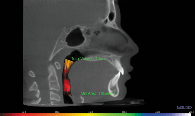

A nine-year-old girl, the oldest of four children, underwent 3D scanning at Dr. Robert Kaspers’ orthodontic office. At first, she appeared to possess a mild skeletal Class II malocclusion with the maxilla forward and mandible retruded. Also, her lower incisors were flared forward. However, an i-CAT 3D scan (in MIP) also showed both condyles were “centered and down” in the glenoid fossa and she had a constricted airway.
Dr. Kaspers (Kaspers Orthodontics in Northbrook, Ill.) decided to utilize a Herbst appliance calibrated to stimulate growth of the mandible to correct the skeletal Class II asymmetry and improve her constricted airway. After treatment, her total volume increased to 14.4 cc from 7.1 cc.
“I am an engineer so I particularly enjoyed having Dr. Kaspers’ diagnosis and treatment plan visualized through 3D imaging,” the patient’s mother said. “When he told me my daughter had issues with her jaw, he was able to show me why. It was especially interesting to see before-and-after images. The orthodontist can tell you the problem is fixed, but it’s nice to actually see it. Of course, I trust Dr. Kaspers, but, as a parent, it is nice to have that visual confirmation.”
Related reading: Applying i-CAT FLX in your practice
When the second child in the family, now 12 years old, was old enough for his orthodontic consultation, his 3D scan also showed he had a constricted airway. With the family history aligned toward airway constriction, the parents decided to have their eight-year-old twin boys checked to address any issues early.
One twin’s 3D imaging showed he had an extremely constricted airway so Dr. Kaspers will likely utilize the Herbst appliance twice during treatment (once during the initial phase and once during the full comprehensive orthodontic phase). The other twin’s imaging revealed what the family nicknamed a “PAC-MAN tooth” because it is positioned completely sideways.
Continue to page two for more...
“We had no idea it was there,” the mother said. “We are now monitoring that tooth, and, in time, Dr. Kaspers will put a chain on it to rotate and twist it into position so the root of the tooth will actually grow into the bone like it is supposed to. Without that orthodontic treatment, that tooth would just have to be extracted.”
Knowing the exact position of that tooth will facilitate calculations to precisely move it into the proper place.
“Dr. Kaspers was able to obtain data about that tooth from the scan so he will be able to fix it,” the patient’s mother said.
During one orthodontic visit, Dr. Kaspers told the mother her daughter would not snore because her airway was no longer constricted. She mentioned her husband also snored badly and asked him to take examine him, as well.
Related reading: How i-CAT FLX is advancing diagnosis and treatment planning
Her husband was about to start wearing a sleep appliance made by his general dentist. Dr. Kaspers took an i-CAT scan to verify the airway was open when the appliance is seated, which it was. He now wears his retainers to keep his airway open at night so he breathes better.
The patients’ mother understands the importance of having all possible details of comprehensive orthodontic treatment provided.
“I appreciate that Dr. Kaspers has the technology to assist him in dealing with health issues that may happen to my kids as they grow older,” she said.
She feels other parents looking for an orthodontist may want to find one who can perform 3D scans.
“For my whole family, that was very important,” she said. “My husband snored and had tension headaches so it’s nice to know my children may not have to deal with those health problems, and Dr. Kaspers identified the condition with i-CAT imaging software. So thank you Dr. Kaspers and i-CAT!”
More on i-CAT FLX and i-CAT FLX MV
i-CAT FLX and i-CAT FLX MV devices construct a three-dimensional model from images taken during a rotational X-ray sequence. The Imaging Sciences i-CAT scanner is intended to be used whenever a dentist, oral surgeon or other physician needs 3D information of high contrast objects. The system is designed for imaging of TM joint studies, mandible and maxilla for implant planning, sinuses and other areas of the maxillofacial complex.


