How to restore root caries with ACTIVA BioACTIVE-RESTORATIVE material [VIDEO]
Every restorative dentist will be, at some point, faced with a patient that presents with root caries apical to an existing crown margin. The choice is whether to remove the existing crown and additional decay and replace the crown, or to attempt to directly restore the affected area without crown replacement.
Every restorative dentist will be, at some point, faced with a patient that presents with root caries apical to an existing crown margin. The choice is whether to remove the existing crown and additional decay and replace the crown, or to attempt to directly restore the affected area without crown replacement.
Let’s further assume this root caries exists on a strategic abutment of a long span bridge-this compounds the decision, especially if economics does not allow the patient to replace the bridge. What are the options now?
Activa: A Bioactive Restorative Material
One critical aspect that will impact on the ability to restore an area of the tooth or root below an existing restoration will be clinical access to the area.
Often these lesions will be subgingival, so access to restore may require a gingivoplasty, or “mini flap procedure,” to access all the restorative margins. If a flap procedure is not needed for access, use of a soft-tissue dental laser is ideal for this procedure. The wavelength of a diode laser can help with localized hemostasis and is considerably less invasive as far as a zone of necrosis when compared to electrosurgery.
Also, it is difficult to use electrosurgery instrumentation safely around metallic restorations.
The second clinical problem is that the surface of the root is not a very good substrate on which to use resin-bonded dental materials. It is well documented that glass ionomer cements “seal” this type of dentin better than adhesive resins.
The difficulty in using traditional glass ionomers may be in the clinical placement into these difficult to reach areas and then maintaining the field to allow for the self-cure set of the material, which may be minutes. Light-cured resin-modified glass ionomers offer a more realistic approach utilizing command set, but often do not allow long-term diffusion of ions through the resin limiting the “healing effects” of the material.
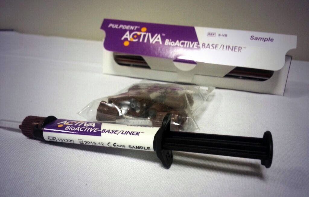
A new RMGI restorative material (ACTIVA BioACTIVE-RESTORATIVE from Pulpdent Corp.) has been shown to allow continual passive diffusion of calcium, phosphate and fluoride ions through the restorative material and to have excellent compressive strength, which is unusual for light-cured RMGI materials.
Activa was chosen to restore this carious lesion on the root of this posterior tooth with a full coverage restoration.
Clinical Case: Root Surface Repair With A Bioactive Restorative Material
Step 1
The patient in Figure 1 of the above slideshow presented with root surface decay apical to existing porcelain to metal crown.
She is in her late 60s and has battled periodontal disease most of her life. Having lost several maxillary posterior teeth and had them replaced with implants, the treatment goal at this time is to maintain her existing restorations as long as possible. In order to gain access to the carious area, a full thickness flap was reflected.
Step 2
A SmartBur II (SS White Burs) round polymer bur is used to carefully excavate the carious area. Thirty-seven percent phosphoric acid is used to “clean the preparation” for five seconds, then rinsed away with copious amounts of water. Air dry, but do not over-desiccate the surface of the dentin.
Step 3
The metal mix tip with metal cannula supplied with ACTIVA BioACTIVE-RESTORATIVE is ideal for access in this area as it can be bent to the appropriate angle to allow for easy and precise delivery of the restorative material to the center of the preparation.
It is important not to overfill the preparation to limit the amount of finishing, since access to the restoration with finishing instrumentation will be difficult. Ideally, just enough ACTIVA Restorative should be placed so that removal of excess or finishing after curing is not needed.
Step 4
Figure 2 in the slideshow shows the ACTIVA Restorative material after placement and light curing. After verifying the accuracy of placement with a bitewing radiograph, the flap will be closed with a silk suture through each side of the interproximal papilla. After one week, the suture is removed.
Step 5
Figure 3 in the slideshow shows a similar type of lesion radiographically on the mesial abutment (tooth No. 31) of a long span bridge. It is financially not feasible for this patient to have the bridge replaced at this time. Rather than section away the pontics (tooth Nos. 29 and 30) from the anterior abutment and extract the distal abutment, it was decided to see if the lesion could be excavated and successfully restored using ACTIVA BioACTIVE-RESTORATIVE.


Step 6
Figure 4 is a post-operative radiograph after placement of ACTIVA Restorative on the root surface of tooth No. 31. There was no pulp exposure during preparation and the patient was told, “we will see how the tooth responds to therapy.” Our hope is that by removing the caries and placing a bioactive restorative material such as ACTIVA Restorative, the ion exchange will help to remineralize any affected dentin remaining and the clinical life of this fixed prosthesis will be extended for an indefinite period of time.
Conclusion
These are cases where teeth with recurrent root caries in difficult to access areas have been restored with a bioactive restorative material in hopes of preventing further tooth loss and extending the life of the existing restorations.
This is one of many clinical examples where a bioactive restorative material, such as ACTIVA BioACTIVE-RESTORATIVE, will prove to be extremely useful in our quest to help patients maintain their dental health.


