How to efficiently restore teeth with a digital scanner [VIDEO]
Accuracy, efficiency and esthetics are likely the top concerns for many dentists when performing crown and bridge work. The accuracy and efficiency considerations are closely tied together, as an inaccurate fit will severely compromise the efficiency of the procedure.
Similarly, efficiency and esthetics can be tied together as well, since achieving the most esthetic results can sometimes mean taking a little extra time. However, the advent of digital impression taking and more advanced milling materials in recent years has resulted in new workflows that allow dentists to achieve an ideal balance between all three factors.
The 3M™ True Definition Scanner is a cornerstone for these new workflows. This digital scanner has demonstrated a fit rate of 99.7 percent, meaning the need for retakes, remakes and adjustments is drastically reduced in many practices.
With crowns and bridges that simply fit the first time, dentists can be assured that their schedules will not be thrown into disarray due to adjustments or remakes.
An additional benefit of working with the 3M True Definition Scanner is its flexibility. The system’s open architecture means its scans can be used with any system that accepts STL files. Dentists can use the scanner as a starting point not only for crowns and bridges, but clear aligners, implant workflows and more.
Crown and bridge work remains the bread-and-butter application for many users of the scanner, however, and rightly so. With a simple scanning procedure that takes just minutes, dentists can quickly capture the data necessary for lab-fabricated or in-office restorations.
Patients benefit as well, with both a more comfortable impression-taking procedure, as well as a seating


appointment that is often shorter than that for a restoration made from a traditional impression.
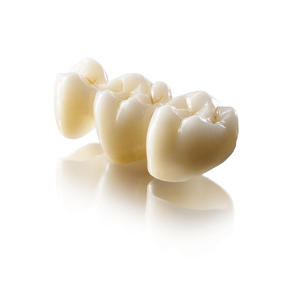
The case illustrated here demonstrates the simple use of the scanner to create a strong and esthetic crown utilizing Lava™ Plus High Translucency Zirconia from 3M ESPE.


Read more about the case presentation and the step-by-step
process on Page 2...
Case Presentation
The patient, a 70-year-old man, had a large MOD amalgam restoration on tooth No. 20 with recurrent caries underneath (Figs. 1-2).
At the time the restoration was initially placed, the patient had chosen it knowing it would serve as a core build-up while he prepared to make an investment in a crown.
A few years had passed, and the patient was now ready to proceed with crown placement.
Step 1. The tooth was prepared routinely for a Lava Plus crown (Fig. 3). The prep in this case involved margins placed at the gingival crest, however, the 3M True Definition scanner can be used with subgingival margins as well.
Step 2. ViscoStat Clear was applied for hemostasis and a size #00 cord was placed for tissue retraction (Fig. 4). Dry angles were used for isolation throughout the scanning process. A fine coat of scanning spray was applied to the teeth (Fig. 5) and the 3M True Definition Scanner was used to capture images of the operative arch, opposing arch (Fig. 6) and bite. The scan process was completed in minutes.
Step 3. The scan was submitted to the lab with a request for a Lava Plus zirconia crown (Fig. 7). This material was selected due to its high compressive strength (important given the patient’s bruxism habit), as well as its esthetics, as the patient was seeking an improvement over the amalgam restoration.
Step 4. A provisional crown was cemented with Tempocem and the patient was given post-op instructions.
Step 5. The patient returned to the office for seating of the final crown (Fig. 8) and the temporary crown was removed. The final crown was tried in the mouth, with the proximal contacts checked first, followed by the margins and occlusion. Once a proper fit was confirmed, the patient was asked for final approval on the shade and overall feel of the crown in the mouth.
Step 6: The preparation was cleaned with glutaraldehyde and the area was isolated. The prep was dried and cotton rolls were placed to help maintain a dry field. 3M™


ESPE™ RelyX™ Luting Plus Automix Cement was dispensed into the crown and the crown was seated on the prep. This resin modified glass ionomer cement is easy to use with a tack light cure option that shortens the cleanup. Excess cement was tack cured for five seconds and then removed, and the patient was instructed to bite on a cotton roll for the remaining set time of five minutes.
Contacts were then flossed and final polishing was performed. The final result in this case was a much more esthetic restoration that can be expected to serve the patient for many years (Figs. 9-10).
Conclusion
The quick scanning and seating processes in this case are very typical for users of the 3M True Definition Scanner. Because the scanner provides the dentist with a chairside view of the data being captured, it is immediately obvious if more attention is needed in any area.
By ensuring an accurate scan is made, the dentist can then be much more confident that the restoration produced in the lab or with an in-office system will fit with minimal adjustments. The ideal combination of accuracy, efficiency and esthetics can be easily achieved with this cutting edge technology.


