Technique: Adhesion dentistry 101
A case study presenting the techniques needed for successful adhesive dentistry.
In 1955, Michael Buonocore, DDS, documented the use of phosphoric acid to improve adhesion of resin to dental enamel1. Since Dr. Buonocore’s observation, adhesion dentistry has played an important role in the evolution of dentistry. Bonding procedures are performed by dentists every day, ranging from bonding orthodontic brackets to delivering stunning porcelain veneers. Because adhesion dentistry is such an indispensable part of the day to day practice of dentistry, a review of its principles will help to refresh the minds and influence the hands of clinicians practicing at every level of experience.


What is adhesion?
In dentistry, adhesion is the chemical and micromechanical union of an adhesive system to enamel, dentin, and a restorative material. Put another way, adhesion is the bond between a tooth and an adhesive, the bond between an adhesive and a resin, and in the case of indirect restorations, the bond between a resin and a restoration. Practical applications of this process include superior retention of restorations, improved performance and durability of certain materials, the ability to affect the color properties under an indirect restoration and low solubility. Clinically, these benefits are tempered by heightened technique sensitivity, increased moisture control and isolation requirements, and shrinkage of resin based materials.
Related article: Bonding resin to tooth structure is still confusing
Optimal adhesion to enamel
Due to its low water content and mineralized crystalline structure, adhesion to etched enamel is very strong and predictable. It is advised that phosphoric acid with a concentration of 32 to 37 percent be applied for 15 seconds on cut enamel, and 30 seconds on uncut enamel.2 Appropriate moisture control measures will prevent enzymes and organic molecules within blood, saliva and crevicular fluid from contaminating the etched enamel. These may include use of various suction devices, a mouth prop, cotton drying aids, retraction cord or a rubber dam. Isolation techniques may include using plastic or metal matrices, wedges and rubber dam isolation.
Up next: See a step-by-step enamel adhesion case


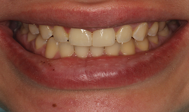
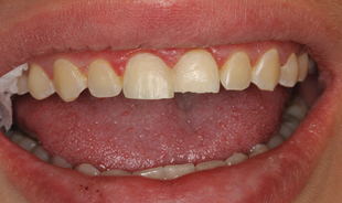
Fig. 1 Pre-Operative
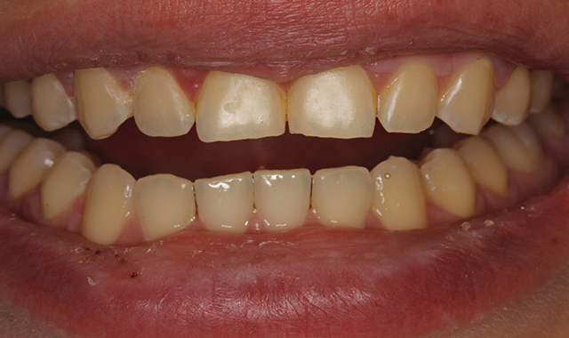
Fig. 1 Pre-Operative
Enamel adhesion case
A post-orthodontic direct composite case is an example of enamel bonding, as the discrepancy in the incisal edges are limited to the patient’s enamel (Fig. 1).


Fig. 2 Enamel prepared with coarse diamond
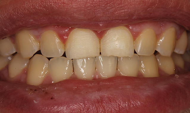
Fig. 2 Enamel prepared with coarse diamond
In this case, the enamel was prepared with a coarse diamond, extending the margin of the bevel far from the actual restoration site in order to improve retention surface area, minimize the optical transition from tooth to resin and move the restorative junction away from the center of the tooth (Fig. 2).

Fig. 3 Etchant appied
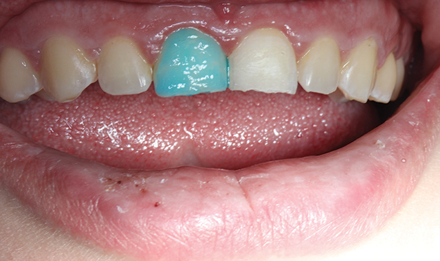
Fig. 3 Etchant appied
It is noteworthy that no dentin was exposed in the preparation. Etchant was applied to the right central incisor for 15 seconds and thoroughly rinsed and dried (Fig. 3).

Fig. 4 Protecting etched enamel
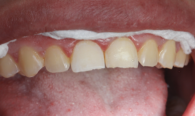
Fig. 4 Protecting etched enamel
Adequately etched enamel will display a frosty white appearance, and should be protected from saliva contamination (Fig. 4).

Fig. 5 Applying adhesive and light cure
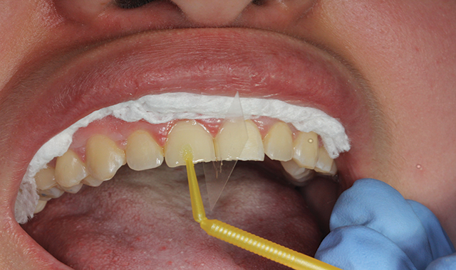
Fig. 5 Applying adhesive and light cure
A mylar strip was used to provide isolation from the adjacent central incisor. All-Bond Universal adhesive was applied in two coats to the etched and visibly moist enamel, followed by gentle air pressure to thin out the adhesive and evaporate the solvent. In this case a wedge was not used, as the patient was not anesthetized. The adhesive was light-cured for a minimum of 10 seconds (Fig. 5).

Fig. 6 Applying composite and light cure
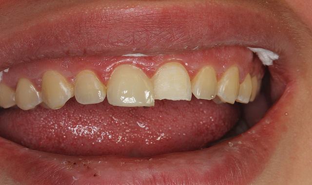
Fig. 6 Applying composite and light cure
Composite resin was applied in two layers and sculpted, with each layer receiving an initial cure through the lingual tooth surface in order to shrink the composite against the facial surface (Fig. 6).


Fig. 7 Roughing in
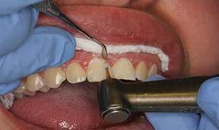

Fig. 8 Primary facial anatomy accomplished
The final contour of the restoration was accomplished using a carbide finishing bur to blend in the margins, while an instrument was used to protect the gingiva from trauma (Fig. 7). Primary facial anatomy was also established at this stage (Fig. 8).

Fig. 9 Polished surface
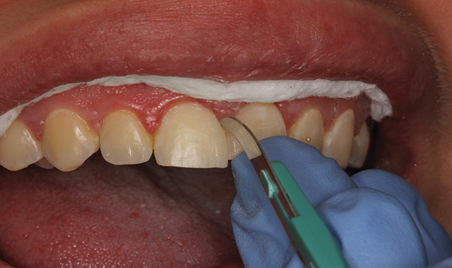
Fig. 9 Polished surface
A #12 scalpel was used to remove any interproximal flash and define the facial embrasure (Fig. 9).

Fig. 10 Polished surface
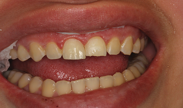
Fig. 10 Polished surface
Brown and green composite finishing rubber points were used to attain a polished surface (Fig. 10).
Fig. 11 Final result
Fig. 11 Final result
The same procedures were used for the left central incisor, and the patient was satisfied with the final result (Fig. 11).
Conclusion and looking forward
Adhesive dentistry is a staple of most dental practices today, and will continue to play a role in the dentistry of tomorrow. In future articles, we will review adhesion topics including best practices for adhesion to dentin, bonding to porcelain and metal, and other areas that will help you provide predictable solutions for your patients.
1 Buonocore MG. A simple method of increasing the adhesion of acrylic filling materials to enamel surfaces. J Dent Res. 0955;34(6):849-853.
2 Suh, Byoung In. Principles of Adhesion Dentistry: A Theoretical and Clinical Guide for Dentists. Newtown, PA: AEGIS Communications, 2013.


