Top Features of the ProMax 3D Imaging System from Planmeca
The ProMax 3D imaging unit from Planmeca features a broad range of imaging views that make it well-suited for diagnosis, planning, and treatments including oral surgery, orthodontics, and implants.
The ProMax 3D imaging unit from Planmeca features a broad range of imaging views that make it well-suited for diagnosis, planning, and treatments including oral surgery, orthodontics, and implants.
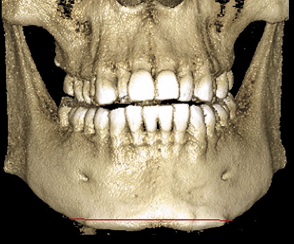
Its three imaging planes make it easier to locate and trace the mandibular nerve canal, providing the dentist with more confidence when planning dental implants.
MULTIPLE IMAGE SIZES: The 3D 8 x 8 cm image size is optimal for most diagnostic applications that require the whole dentition, mandible, and maxilla in the same volume. The 8 x 5 cm volume can be used for single views of the mandible or maxilla, lowering the radiation by almost 40%. The small 4 x 5 cm volume is intended for molar area studies or planning 3rd molar extractions. The volumes can be blended together to generate an image up to 14.4 cm x 10.5 cm x 8 cm horizontally and 8 cm x 8 cm x 13 cm vertically. Volume reduction to lower radiation exposure offers true 2D panoramics and cephalometrics, eliminating the need for multiple imaging units.
IT'S UPGRADABLE: SCARA (Selectively Compliant Articulated Robotic Arm) technology allows existing ProMax users to upgrade an already existing unit, which saves money on new capital equipment investments. This patented technology also enables free geometry based on image formation and can produce any movement pattern required; this allows accurate and reliable volume positioning, volume diameter adjustment, and a reduction in radiation exposure.


QUALITY IMAGES: Panoramic images are taken with the same flat-panel sensor as the 3D images.
ADDED PLUS: SmartPan can be added to the 3D unit, creating 10 layers to reduce risk of patient positioning errors while taking the image. The user can browse between the layers in Romexis Imaging Software.
Watch a Video to Learn More...
Now Playing: Jim Hooper, District Sales Manager for Planmeca, discusses the benefits of the ProMax imaging system at last year's Chicago Dental Society's Midwinter Meeting.