Top technologies for caries and cancer detection
When it comes to technology and the integration of it, there are many ways to skin the “Technology Cat.” For many, it might mean integrating computers into your daily clinical routine. Or it might mean finally embracing digital radiography and adding that component to your office armamentarium.


When it comes to technology and the integration of it, there are many ways to skin the “Technology Cat.” For many, it might mean integrating computers into your daily clinical routine. Or it might mean finally embracing digital radiography and adding that component to your office armamentarium.
However for this article, let’s take a look at a couple of technologies that can literally improve your patients’ quality of life or even save a life. These technologies are digital caries detection and oral cancer screening.
Digital caries detection
Digital caries detection can be divided into two types. These are “probe based” and ”visual based.” The probe-based device is the Kavo Diagnodent. I consider this the 800-pound gorilla in this product category. This is mainly because it has been around for about 15 years and because of that longevity, almost everyone in dentistry has heard of it and is at least familiar with it. This is also probably because it is the only probe-based device still available for purchase.
It uses a 655 nanometer diode laser. At this wavelength of light, caries will glow or fluoresce. The Diagnodent probe is lightly traced around clean pits and fissures similar to the way we used to use explorers. When it detects the fluorescence signature of caries, the device emits a tone and gives you a numeric score. The tone increases in pitch as it detects larger carious lesions and corresponds with a higher numeric score.
The device is very accurate, but it does have some limitations. It does not work around any restorations or sealants as they give a false positive result. Also the teeth must be cleaned of plaque by using a prophy brush or some type of particle delivery such as air abrasion or a Cavijet as plaque will also give a false positive.
The other limitation is that it is difficult to completely replicate scores from one prophy appointment to the next in case you want to monitor a small lesion and see if it increases in size over time.
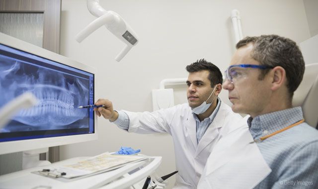
The other category is the devices that make up the visual-based side of the equation. These are devices such as , Acteon’s SoproLIFE and SoproCARE, DEXIS’s CariVu and the Canary System.
Check out this interview with Boaz Munnerlyn of DEXIS on DEXIS CariVu and why it won a Best of Class award:
All of these devices use some type of camera system to display tooth images on the operatory computer. Spectra, as well as SoproLIFE and SoproCARE, use special light wavelengths to display caries fluorescence. CariVu uses invisible infrared light that is seen by the device and displays the tooth on the screen in a view similar to a bitewing radiograph, but uses transillumination instead of radiation. The Canary System uses a laser to image the tooth and display caries on the monitor with penetration of the tooth up to 5 mm.
The advantages of these systems are the ability to easily replicate images from one appointment to the next and the ability to show patients an actual photo of their decay. This gives much more credence to the process and helps the patient understand their clinical situation. They are also very good devices for monitoring lesions in case something small is detected and the doctor wishes to try remineralization therapy.
The images can be stored in your practice software so that they can be monitored from one appointment to the next. Since it is very easy to duplicate these images on separate appointments, monitoring of small areas is easy and accurate.
Oral cancer screening
One of the most important prophylactic services we can render is oral cancer screening. As one of the causative factors is HPV 16, we are seeing the age of affected individuals drop from middle age into the 20s.
Fluorescence can also be used in this situation as well. By using specialized wavelengths of light, oral tissues can be examined and screened. These devices do not connect to the operatory computer so the screening relies on the doctor’s eyes alone.
The devices are LED Dental's Velscope VX, Dentaleze’s Identafi and Dentlight’s Dental Oral Exam kit (DOE for short).
When viewed by these devices, healthy tissue has the appearance of a green apple while injured or cancerous tissue appears black. The Velscope VX and the Dental Oral Exam system both use a single wavelength of light for screening. The Identafi uses two wavelengths for screening. The first is the standard tissue fluorescence and the second is an amber wavelength that penetrates the tissue much deeper. When a dark area is discovered, this amber wavelength will penetrate deeper and show vascularity of the lesion if it exists. Since cancerous tissue has high vascularity, this helps to confirm the presence of neoplastic tissue.
The Velscope VX has a special attachment that allows for a touchscreen iPod to be connected to the device, which can be used to take a photo. The photo can then be sent to the appropriate referral office so that the doctor can see exactly where the lesion is located.
We do fluorescence screening on all patients 17 or older and I am considering dropping that age to 15. The exam process takes just about two minutes and is identical to the screening you would do with white light.
We do not charge for this screening although I know many offices that do. I include it as a value added service for patients and sometimes receive referrals based on the screening.
Wrapping it up
All of these devices are able to allow the doctor to receive much more data than ever before. Being able to diagnose and or monitor changes in the mouth is something that allows the office to be much more conservative in diagnosis, monitoring and treatment.
Patients have come to expect leading edge technologies from my office and using these devices helps reinforce that expectation. They are also grateful for the services since it allows us to treat and screen. We share images and information with patients at every continuing care appointment and it fits well with our patient education platform.
So there you have it. Using these devices will help you be a better diagnostician as well as more conservative with treatment. The devices are not prohibitively expensive and work just as advertised. I highly recommend you explore t


