How to bridge the gap between traditional and digital impressions
A new impression material that might make the jump to digital dentistry a little less jarring. While more dentists are utilizing digital impressions in their offices and some are milling restorations on site, there are still many, like me, who prefer conventional impressions.


While more dentists are utilizing digital impressions in their offices and some are milling restorations on site, there are still many, like me, who prefer conventional impressions.
By using an impression material that can be scanned by the laboratory or scanned in the dental office and the image sent to the laboratory, many advantages of digital technology can be gained.
A new scannable impression material
BONASCAN by DMP is a new polyvinal siloxane impression material that can be digitally scanned. A conventionally fabricated restoration can be fabricated by pouring up the impression and making a hard model, but if the dentist wants to use the digital feature they have two options:
1. Speed up the restoration process as the lab would get the file the same day and can start working on the case. This technique can:
- Improve communication with the lab as a discussion can be had while both looking at the digital file.
- Improve efficiencies for the lab, saving them time and money as they can skip the step of pouring a model and using a powder or spray to scan the model into their system.
- Allow the use of desktop digital scanners, which are less expensive, smaller and easier to use than CAD/CAM systems.
- Allow the dentists to work with a restorative material, not limited by the CAD/CAM software programs that only work with certain milling machines.
- Eliminate the potential for impression distortion during the shipping, handling and model-pouring stages, leading to better clinical outcomes.
2. Take the traditional impression, send it to the lab, ask the lab to directly scan the impression and have them send you the file. This technique can:
- Improve efficiencies for the lab, saving them time and money as they can skip the step of pouring a model and using a powder or spray to scan the model into their system.
- Eliminate the potential for distortion during the model-pouring step, leading to better clinical outcomes.
- Allow the dentist to get a digital file for their records.
- Allow the dentist to focus on patient care and not worry about operating and maintaining CAD/CAM systems.
- Allow the dentist to participate in digital technology with no upfront investment in digital equipment.
Clinical example
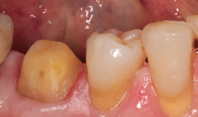
Figures 1 and 2 show the facial and occlusal views of a madibular molar that I prepared for a full crown.
In Figure 3, you can see a close up view of the final impression taken with BONASCAN impression material. A light body was used to express around the margins and a heavy body was placed in the impression tray and seated directly over the light body. After five minutes, the impression was removed and evaluated. A provisional crown was fabricated and the patient was reappointed for seating.


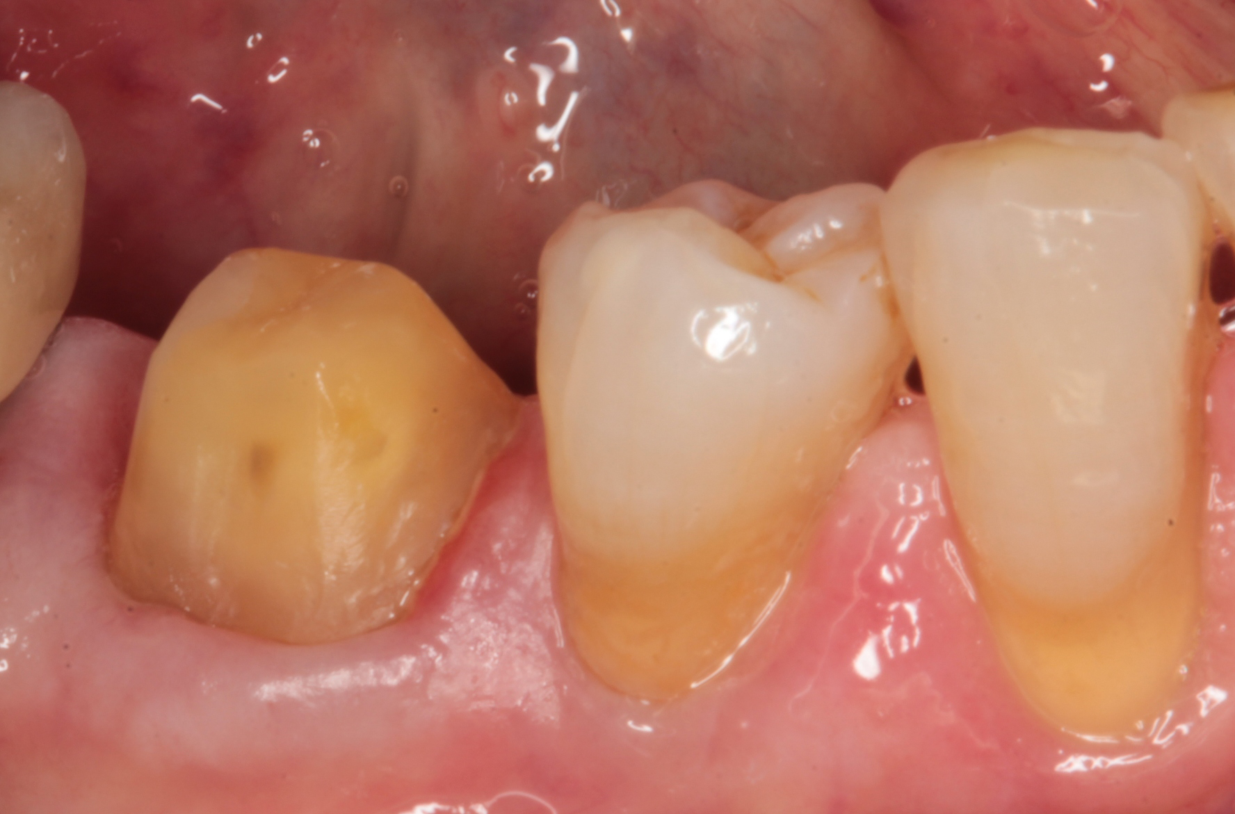

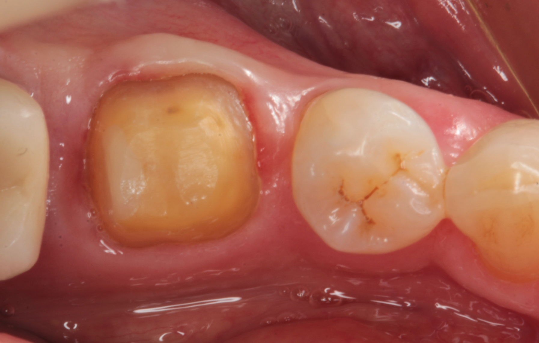


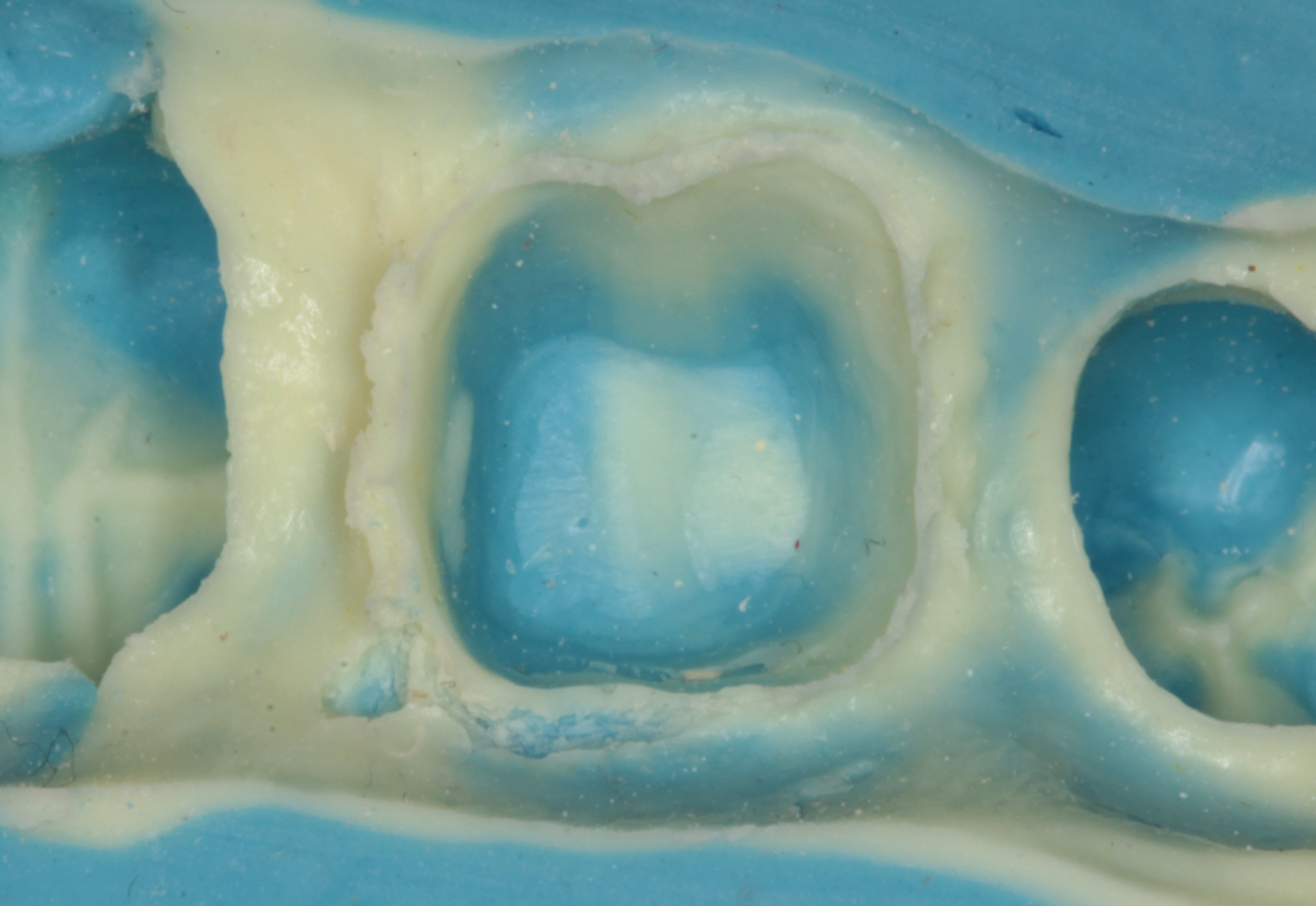
Fig. 1 Fig. 2 Fig. 3
The impression was sent to the laboratory (Peter Kouvaris, New York City) and a lithium disylicate (e.max, Ivoclar Vivadent) crown was prescribed using digital technology. In the lab, Peter scanned the impression and developed the digital information (Fig. 4).


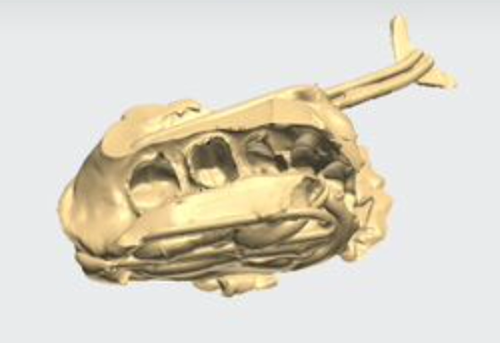


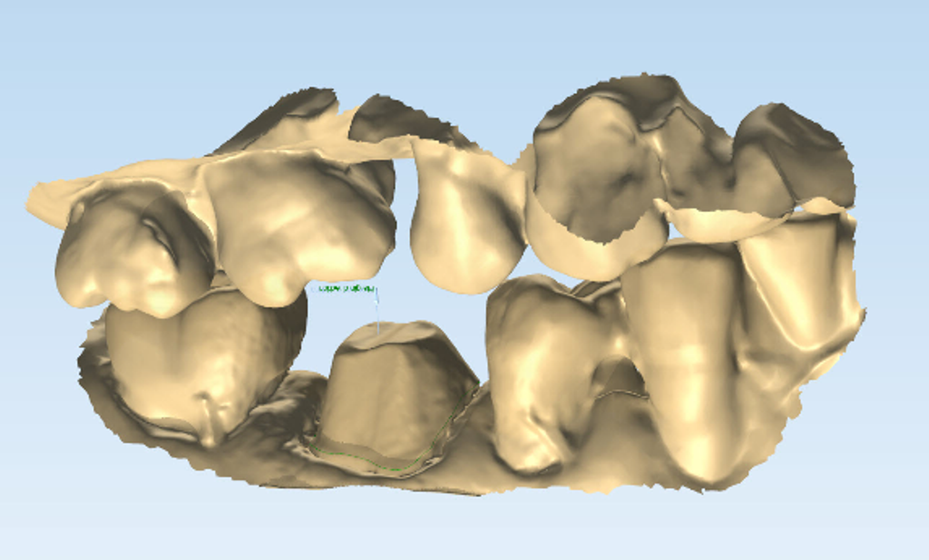


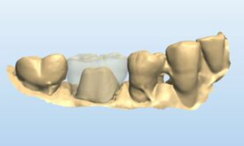
Fig. 4 Fig. 5 Fig. 6
Figure 5 shows a digital image of the teeth in occlusion. Peter designed the crown using digital technology.
The crown can be seen designed on the model from the facial view in Figure 6 and occlusal view in Figure 7. The crown was milled and Peter added surface staining for esthetics. The final restoration can be seen on a mirrored surface in Figure 8.


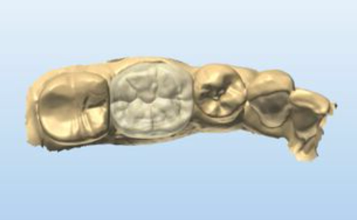


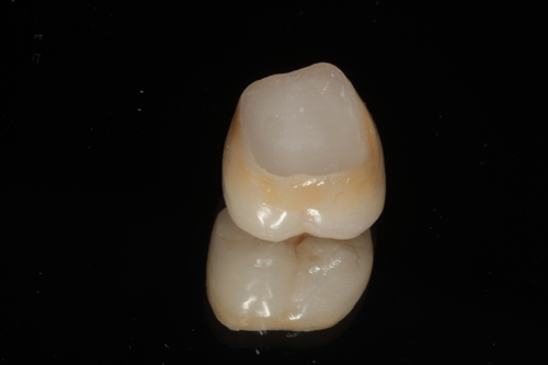
Fig. 7 Fig. 8
While not a necessary step, Peter fabricated a 3D-printed model of the teeth to verify the fit and occlusion. In Figure 9 the printed model is shown and the crown is seated on the model in Figure 10.
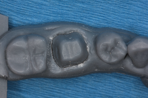


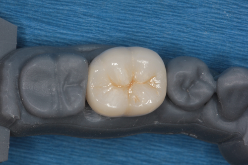
Fig. 9 Fig. 10
The crown and printed model were returned to my office. At the seating appointment, the provisional crown was removed and the final crown was tried in. Contacts, margins and occlusion were evaluated and no adjustments were needed. The crown was bonded to place using a self-adhesive resin cement (Fusion-ZR Dual Cure Resin Cement, Taub Products). It can be seen in place from the facial view in Figure 11 and the occlusal view in Figure 12. The radiograph in Figure 13 shows the excellent fit of the digitally fabricated crown.

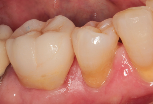


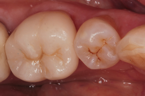


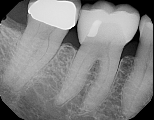
Fig. 11 Fig. 12 Fig. 13
Conclusion
For the dentist who would like to take advantage of digital technology to produce restorations but prefers conventional impressions, a scannable impression material can fill the gap in technology.


