Why you should switch to digital sensors
Dentists all over are making the switch - so what’s holding you back?

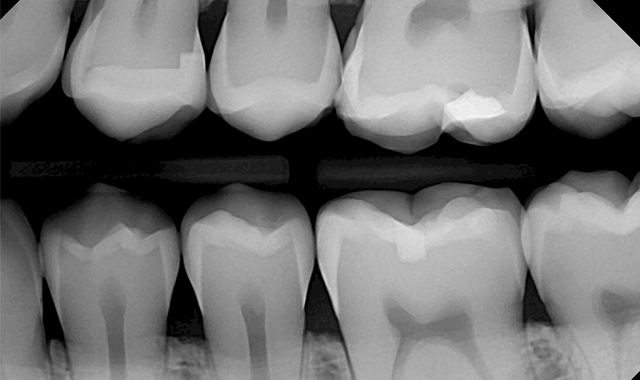

In dentistry, radiographic images play a major role in diagnosis. In order to arrive at an accurate diagnosis, it is critical to have a high quality radiographic image. And what is the best way to achieve high-quality images in this day and age? I believe that most dentists will agree that digital sensors are the answer.
I used film from 2000 to 2016 mainly because that is what I had and was familiar with. I was getting acceptable images, but I decided to switch to sensors a couple of years ago for a few reasons.
First, the images appear instantly with excellent quality. Second, digital radiography has largely become the standard of care. Dental patients these days generally expect their dentist to use digital imaging. Third, digital radiography allows us to deliver a lower dose of radiation per exposure, which is important in our efforts to follow the ALARA principle.
In addition to these important reasons, digital radiography allows us to do away with the various issues associated with developer chemicals and the developer itself.
After realizing that I needed to go digital, I started looking around at different sensors and comparing a few different brands. When I finally found what I was looking for, it was time to make the switch. Much to my surprise, the transition was much easier than I had expected.
Techniques used with digital sensors are quite similar to those used with film-based radiography. Any adaptations for using digital sensors are quite minor. I had always used RINN® positioners (Dentsply Sirona) for positioning film, and I still use positioners with my sensors. A good quality positioner is vital to obtaining a good image. Simply lining up the X-ray head correctly to the positioning arm and positioning ring will assure that angulation is correct and facilitates the capture of an optimal image.
However, it is important to mention that there is more to achieving good images than simply having good technique. If you really want to obtain good quality images, you will also need to use high-quality sensors. After doing some research and trying a few sensors, I found that the DentiMax Dream Sensor delivered excellent image quality.
The images were as good or better than the competitors’ images. Surprisingly, the price was much lower than the other sensors I had sampled. I was very happy to find a sensor that delivered such excellent images at such a reasonable price.
Certainly, one of the most challenging parts of switching to sensors is getting familiar with the software. To help us with this, DentiMax provided training, which got us off to a great start. We were also able to have some time with a trainer after a week or two of purchasing the sensors to allow us to address any questions we had. We found this to be very helpful.
I feel like DentiMax has been very interested in assuring our success with their sensors. If you are switching to sensors, having good training and tech support available is definitely important. I have been impressed with the trainers and support staff at DentiMax; they have been easy to work with and kind and helpful.
Once you learn how to get the most out of your sensors and become familiar with the software, the results are just phenomenal. With sensors, I am getting higher quality images than with film, which allows me to detect pathology better. If we have to retake an image, sensors save us a lot of time because I can see the image immediately and retake the image within seconds.
Besides that, we do not have to mount the film like we did with film X-rays. With the tools that software like DentiMax has, you can use a preset mount that the images flow directly into. This is a big time saver for my assistant.
And let us not forget about the advantages for the patient. The most obvious one is the lowered dose of radiation with each radiograph.
But another benefit is that I am able to show the images to the patient instantly. I have the images on a computer screen, which can be positioned very close to the patient. I can enlarge the images or make other modifications to allow the patient to better visualize any issues that I need to discuss with him or her. This helps the patient to feel more comfortable and confident in my diagnosis and treatment.
If you are considering making the switch from film to digital, I would recommend trying out some sensors first. Find a company that can give you a demo before you buy. And if you are hesitant about making the switch, well, don’t be! It’s not as difficult as you may think. In my experience, the transition was quite seamless, and we were able to get up to speed very quickly. Make the switch. You will be glad you did.


