Tips, Tricks, and Best Practices for Spotting Root Fractures
Tooth cracks fractures can be hard to spot, but it’s important that they’re treated. Here is some key insight on root fractures from endodontic professionals.


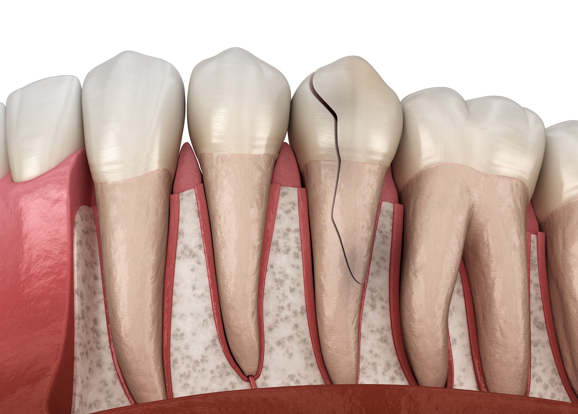
Tips, Tricks, and Best Practices for Spotting Root Fractures: © Alex Mit - stock.adobe.com.
The Grand Canyon is 1 mile deep, 18 miles wide, and 277 miles long. Tooth fractures (even the widest) are about 112 million times smaller than the Grand Canyon, but at 250 µm, those pose a real risk to patients. Finding those cracks and fractures can be tricky, even for the most seasoned endodontist and takes a combination of tools, the endodontist’s skills, and their years of experience.
Determination
No patient wants to hear that they have a cracked or fractured tooth. However, those fissures are not uncommon. Their specific characteristics are when they get worrisome.
“Cracks and fractures are probably the hardest thing that I deal with and diagnose every single day,” Ryan M. Walsh, DDS, MS, Diplomate, American Board of Endodontics and endodontist at Advanced Endodontics of Texas says. “I tell my patients that cracks and fractures are like Baskin Robbins ice cream–they come in a bunch of different flavors. How long have they been there? How deep are they? But it’s my job to figure out which flavor it is and is this something that we need to treat or just watch? I guarantee right now, if you were to sit in my dental chair, I’d find cracks and fractures all over your teeth, and you would also find those on mine. But when do they become an issue of pathology? When do they become an issue of symptoms? To me, once a crack or a fracture becomes symptomatic, that’s where I draw a line in the sand of, ‘It’s time to treat.’
“But how do we treat? That’s a bigger question,” he continues. “Our studies show that that 80% of crown fractures–if they’re caught early and they’re the fracture is maintained on the surface of the tooth–it only needs a crown. Only 20% of the time do they have to come see me because of that fracture. So, I think if you’re giving me 80% odds to go to Vegas, I’ll take that all day long. By and large, we can treat these very conservatively, and 20% of the time they need root canals. But still, that’s a very predictable, long-term success with that tooth. It’s only when it’s ignored and/or patients have been symptomatic for a very long time do we really get concerned about it progressing from a crown fracture to a root fracture. And once it’s made that progression from crown to root, the long-term prognosis drops off the cliff.”
Fractures, once discovered, must be assessed to determine just how serious they are.
“As an endodontist, the more important question that my partner and I are often pressed to answer is not only is there a fracture present, but does that fracture extend onto the root structure?” Rebekah Pryles, DMD, Diplomate of the American Board of Endodontists and endodontist in White River Junction, Vermont says. “It’s an imperative question that we answer, because we know that fractures that extend onto root structure are associated with bone loss and loss of the tooth.”
Establishing the crack’s severity will determine the best course of action.
“The way I describe a fracture to a patient is like splitting up firewood,” Dr Walsh says. “The more pressure you place down, the farther it opens that fracture. That’s what you feel when you bite. So, most patients will have sensitivity to bite. That is normal. I kind of draw the line in the sand of how long has it been going on? And equally as important, is it sensitive to cold or heat? If it’s sensitive to cold or heat and that lingers beyond–let’s just say an arbitrary number like 10 seconds–that tells me there’s more inflammation inside that pulp than really meets the eye. At that point I’d tell the patient, ‘If I go by the book, you don’t technically need a root canal now, but the chance of you needing one based on these symptoms after you get a brand new crown is really high.’ In these cases, I think it’s best to go ahead and do it prophylactically, because at this point, there’s no bacteria involved. The prognosis of your tooth is the best it could possibly be with a root canal, and we’re saving you pain and energy down the road. Plus, I don’t have to drill through a brand new crown that you just spent a thousand bucks on.”
Tools
It should come as a shock to no one that technology continues to improve our lives and our work in particular–and endodontics are no different.
“For us, one of the single most important tools at our disposal is use of the cone beam,” Dr Pryles says. “Cone beam CT imaging has really caused a shift in endodontics. For most patients, cone beams aren’t going to show fracture lines, themselves, but rather those characteristic bony changes associated with fracture. Historically, before we had cone beams available, we were having to do a lot of exploratory treatment–open up the tooth and see if that crack goes down onto root structure. And now, if we’re seeing some of those characteristic bony patterns in the presence of clinically detectable fracture lines, we know we’re dealing with root fracture from the get-go. And we can send that patient for extraction, which is the appropriate treatment at that point.”
There was a time when x-ray technology revolutionized dentistry. And while it is still a mainstay in dental practices, it’s not the best tool for endodontists.
“If I can find it on an x-ray, it’s too late,” Dr Walsh says. “Oftentimes it’s the patient’s symptoms and then a visual identification. If I’m seeing a big fracture on a CT or I’m seeing radiographic evidence, usually that’s too late.”
Missteps
What are the common mistakes that clinicians tend to make when assessing cracks and fractures?
“Something that I see a lot in the endodontic social media world is that it would be easy to assume that every J-shaped lesion that we see on periapical images or on cone beams are caused by fractures,” Dr Pryles says. “But we know that that’s not the case. We know that things like periapical infections can cause really similar radiographic pathology to fractures. And so, I would argue that an easy mistake to make would be the overdiagnosis of fracture.”
“There is a tendency to undertreat, by a landslide, because oftentimes we can’t visually see it, so a very common chief complaint is the patient says, ‘Every once in a while, I bite and I get this zinger, but it doesn’t happen every time. It only happens once in a while.’ So, the dentist looks, does their evaluation, makes a radiograph, and they don’t see anything. Just because you can’t see it doesn’t mean something’s not wrong. If the patient’s feeling these symptoms, then it warrants further investigation. I can use transillumination. I can use a Tooth Slooth or a bite stick to figure out where that fracture’s coming from. Once I identify the fracture, then I can say, ‘Should it be just crowned? Should we do a root canal and crown it?’”
Pro Tips
Drs Walsh and Pryles offer some of their best tips for finding cracks and fractures:
“Transilluminate and use the Tooth Slooth or the bite stick on every single cusp,” Dr Walsh says. “They’re inexpensive tools that almost everybody has in their office. They can help expose that fracture and help us diagnose it early to allow for more conservative treatment.
“The other thing would be, listen to the patient,” he continues. “If they’re having occasional or sporadic pain on biting, a fracture should move up very quickly in your differential diagnosis.”
Ultimately, it is a combination of all the endodontist’s skills and tools that help ferret out fractures.
“Like any dentist out there, using all of the information you have at your disposal – both your clinical exam findings coupled with the radiographic findings – to accurately diagnose is really the best way to pick up those fracture lines,” Dr Pryles adds. “Clinically, we want to probe all of our teeth, especially those that are coming in for endodontic evaluations to assess for isolated, deep pockets. We want to look at the position of sinus tracts. We know that most sinus tracts, if they’re just of simple endodontic origin, are going to be located apically, near the debris apices, whereas those associated with fractures or endo-perio pathology are often more coronally located. And we want to put that clinical information together with what we’re seeing on our imaging–whether it’s 2D or 3D–to accurately diagnose. We can’t diagnose from a radiograph alone. We can’t diagnose from clinical findings alone. It’s really a matter of putting it all together and being systematic about what we’re looking for when we’re talking about fracture diagnosis.”
A canyon that is a mile deep, 18 miles wide, and 277 miles long is pretty easy to spot. It’s trickier for endodontists, however, to find those fractures that are only 250 µm wide. But it’s critical because even something so small can lead to big problems for patients.


