Researchers develop AI model to detect mandibular canals
Researchers at the Finnish Center for Artificial Intelligence (FACI), Tampere University Hospital, Planmeca, and the Alan Turing Institute have developed a new model that accurately and automatically shows the exact location of mandibular canals.

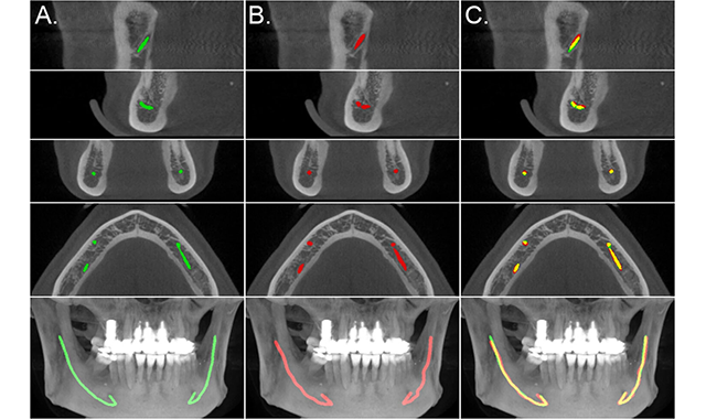

Researchers have developed a new model that can automatically localize the mandibular canals for dental implant operations.
In order to plan an implant operation, dentists must use X-ray and computer tomography (CT) to detect the mandibular canals manually. This process can be often laborious and time-consuming. Fortunately, there may be a solution on the horizon.
Researchers at the Finnish Center for Artificial Intelligence (FCAI), Tampere University Hospital, Planmeca, and the Alan Turing Institute have developed a new model that accurately and automatically shows the exact location of mandibular canals. The researchers trained the model by using a dataset consisting of 3D cone beam CT (CBCT) scans.
The model is based on a fully convolutional architecture, which makes it as fast and data-efficient as possible, the FCAI said in a press release. Based on research results, this type of deep learning model can localize the mandibular canals highly accurately and is said to be able to surpass statistical shape models.
In simple cases, such as when the patient does not have any special conditions, the model is just as accurate as a human specialist, according to the study.
“In more complex cases, one may need to adjust the estimate, so we are not yet talking about a fully stand-alone system,” Joel Jaskari, a doctoral candidate and the first author of the research paper, said.
“The aim of this research work is not, however, to replace radiologists but to make their job faster and more efficient so that they will have time to focus on the most complex cases,” Professor Kimmo Kaski added.
Dental manufacturing company Planmeca has collaborated with the FCAI on this study. Currently, the company is integrating the presented model into its dedicated software for use with its Planmeca 3D tomography equipment.
The results of the study were published in Nature Scientific Reports.


