Panoramic X-rays: Taking a Wide View on Radiography
Radiography helps you diagnose problems and diseases. We talk to experts about why it is essential to incorporate panoramic X-rays into your workflow.


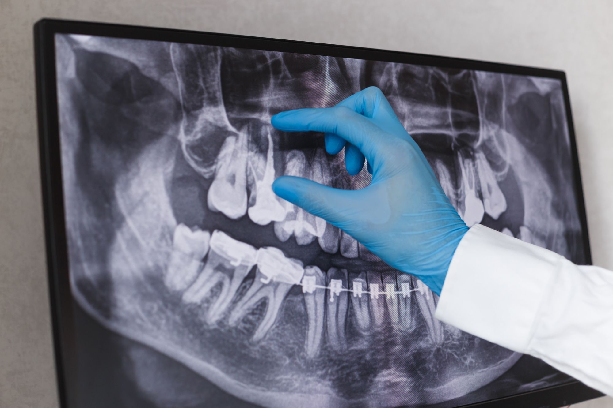
Panoramic X-rays: Taking a Wide View on Radiography. Image courtesy of vladdeep/stock.adobe.com.
Panoramic X-rays can capture the entire oral cavity in a single, two-dimensional (2D) image. There are a variety of clinical benefits, the patient care benefits, and considerations a dental practice should remember before investing in a panoramic X-ray system.
Panoramic X-rays show dental professionals many things about a patient's mouth. But perhaps the most significant benefit is that it shows the jaws and the teeth in 1 image, which allows dentists to understand what is happening in the patient's oral health.
For example, these images show the effects of advanced periodontal disease, cysts in the bones, jaw tumors and oral cancer, impacted teeth and wisdom teeth, temporomandibular (TMJ) joint disorders, and sinusitis.1 In addition, dental professionals might use these images to assess dental restorations or for dental surgery prep.2 Some dental professionals use panoramic X-rays to see infections or as a pre-treatment image for implants and dentures treatment planning.3
Dental teams use panoramic X-rays for many indications. These include unerupted third molars, assessments for orthodontics, observing tooth development, detecting developmental abnormalities, or assessing trauma and significant lesions. Regarding anatomical variations, there are a few that dentists might find on their images, like bifid mandibular or retromolar canal. A dentist might also detect alterations like a calcified stylohyoid complex (hardening of ligaments), arterial calcifications (mineral buildups in the arteries), phleboliths (calcium in veins), sialolithiasis (hard stones in the salivary glands), or tonsilloliths (hard bits tucked into the tonsils). The panoramic X-ray is usually the initial radiology in these cases, and the dentist uses it to determine if a more detailed exam is necessary (i.e., a Cone Beam Computed Tomography or CBCT, scan) for diagnosis and treatment or if the patient needs a referral to another healthcare professional or dental specialist.4
However, they are not a great tool for clinical situations that require details. For example, a panoramic X-ray would not be the best option for carious lesions, seeing the alveolar crests, the level of root canal filling, and gum disease or periapical lesions. These indications require more definition than Panoramic X-rays can deliver.4
The Clinical Benefits of Panoramic X-Rays
Mark Greenwood, Vice President of Imaging Product Development at Midmark®, describes the panoramic X-ray as an excellent survey instrument to review a patient's overall dental health. These images cover a wide variety of dental anatomy and serve as a good starting point for looking at the patient. For example, not only can the clinician see the mandible and the maxilla in one image, but they can also see problems with the tissue like tumors, cysts, and the effects of gum disease—and all types of things you would not see in an intraoral image.
"Intraoral images are high-resolution images that give you small images of each tooth. The panoramic gives you a nice survey of the dental arch," Greenwood says. "It's a great overall survey tool for looking at the patient."
Greenwood says you can also look at specific problems with a panoramic X-ray. For example, clinicians can see impacted teeth and other teeth positioning issues across the mandible and the maxilla. In addition, clinicians can diagnose advanced periodontal disease and tumors, 2 things that would not be in a place they can see with an intraoral image.
"An intraoral image doesn't go much above or below the root tips," Greenwood says. "Panoramic is good for looking at TMJ issues because they include the condyles of the TMJ. They also go into the lower sinus so you can see things like sinusitis and other issues with the sinus."
However, Greenwood thinks the overall survey use case makes it even more helpful as something to have for your patients over time. The number of panoramic X-rays you can take varies by insurance companies, but a general time frame is every couple of years. So, the dental team can compare images over the years and see any changes.
"It's also a good communication tool between the dentist and the patient. In an intraoral image, patients don't always understand what the dentist is showing them or where it is in the mouth. It's only a portion of the tooth, and sometimes it doesn't always do the trick," Greenwood says of intraoral image capture. "Most people can look at a panoramic X-ray and say, 'yeah, that's my smile, that's my teeth.', and they understand what it is."
The Patient Benefits of Panoramic X-Rays
From a patient's point of view, Greenwood says that panoramic X-rays are more comfortable than bite-wing X-rays. Midmark does market research related to X-rays and patient experience at the dentist. It shows that patients do not like the sensor shoved into their mouths. Patients with smaller mouths or a strong gag reflex find it especially difficult.
Another benefit Greenwood and the team at Midmark learned through their research is that dental schools that had a clinic used the panoramic X-ray more often during COVID. There were fewer infection control concerns since the panoramic X-ray was extraoral and did not go in and out of the patient's mouth.
Also, Greenwood says a panoramic is efficient from an X-ray radiation perspective. The beam that is delivered is very collimated. The thin, narrow beam has less radiation to the patient than a full-mouth series or a CBCT.
"They also take less time," Greenwood says. "The panoramic is a 20-second scan. So, it's a shorter time frame than a full-mouth series, which takes several minutes for an assistant to go through."
What to Consider Before Investing in a Panoramic X-Ray System
Greenwood thinks dental practices that want to invest in a panoramic X-ray system should consider a few things, including:
- Where to put the unit: Greenwood says floor space is often at a premium in a dental practice. So, it is essential to know how much space the unit needs, the power requirements there, and where you want to put it.
- Whether the practice might want to move to CBCT imaging someday: If so, Greenwood says the dental practice should ensure they can upgrade the unit to include 3D imaging. They should also know what is involved in that upgrade because sometimes they can be extensive. Greenwood says the adoption rate of CBCT is on the rise, making this information essential for a dental practice to know up front before purchasing a panoramic X-ray unit.
- Ease of Use: Advances in digital sensor technology make it easier to capture even more information from panoramic X-rays. For example, Midmark's Extraoral Imaging System (EIOS) system comes with autofocus that brings in more of the image to the focal area.
"When the first unit we installed in the field had that feature, the practice couldn't believe the consistent quality images of the panoramic. They noticed that as an improvement over the older panoramic," Greenwood says.
Greenwood thinks these improvements, like autofocus, can provide improved diagnostic images, especially compared to those panoramic radiology units from 10 or 15 years ago. As a result, panoramic X-rays can be valuable technology units for a dental practice.
"They can provide a lot of functionality and improve patient care," Greenwood says.
References
- Radiological Society of North America (RSNA) and American College of Radiology (ACR) (2022) Panoramic Dental X-ray, Radiologyinfo.org. Accessed October 7, 2022. https://www.radiologyinfo.org/en/info/panoramic-xray
- Watson, S. (2021) How dentist uses Panorex X-rays to find dental disease, Verywell Health. Verywell Health. Accessed October 7, 2022. https://www.verywellhealth.com/panorex-definition-of-panorex-1059431
- What is a panoramic dental X-ray? (2022) www.colgate.com. Colgate-Palmolive Company. Accessed October 7, 2022. https://www.colgate.com/en-us/oral-health/X-rays/what-is-a-panoramic-dental-X-ray#
- de Oliveira Capote, T. S. et al., 2015, 'Panoramic Radiography — Diagnosis of Relevant Structures That Might Compromise Oral and General Health of the Patient', in M. S. Virdi (ed.), Emerging Trends in Oral Health Sciences and Dentistry, IntechOpen, London. doi:10.5772/59260


