The minimally invasive bridge
January 13, 2010 | dentalproductsreport.comWeb ExclusiveThe minimally invasive bridgeThe lingual retained multi staple and groove overlay.by J. Tim Rainey, DDS, MAGD
January 13, 2010 | dentalproductsreport.com
Web Exclusive
The minimally invasive bridge
The lingual retained multi staple and groove overlay.
by J. Tim Rainey, DDS, MAGD

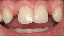

Click on image to review slideshow of Dr. Tainey's cases.
When I started my dental practice in 1971, I was privileged to see examples of precision crown and bridge work from the 1930’s and 40’s that were known as “Pin Ledge Bridges.” These bridges just made sense because the facial of the natural teeth on either side of the pontic remained as they had been when they had erupted, with the pontic presenting as usual in replacing the missing tooth. The advancements of ceramics in the 1960’s and 70’s allowed me to create pontics mimicking natural tooth structure, and with the then new hard and dense non-precious ceramic metals, we closed the loop in esthetics.
According to the World Congress of Minimally Invasive Dentistry, “Minimally invasive dentistry is respecting the health, function, and esthetics of oral tissue by preventing disease from occurring or intercepting its progress with minimal tissue loss.” Early in practice, we rapidly adopted this genuinely minimally invasive approach to anterior crown and bridge as our treatment of choice in replacing missing anterior teeth. With a good ceramist forming the pontic representing the missing tooth, a truly lasting esthetic result could be achieved, with one of our Pin Ledge Bridges done in 1974 still in place, esthetically acceptable, and in function until recently.
Lab matters
Again, these bridges are not only technique sensitive in preparation, but the laboratory must also pay exquisite attention to detail. The metal linguals must be waxed to a proper thickness to accommodate casting, with adjustment to occlusion done in the metal. Recognizing that successful completion of the bridge is an acquired skill for both the dentist and the fabrication laboratory, I would also advise using a skilled laboratory experienced in constructing the first few initial appliances.
Always evolving
The next great advancement in minimally invasive bridge technology was the introduction of Panavia® Crown and Bridge cement, which could form a true bond between the enamel and the non-precious metal substructure of the bridge. This advancement allowed us to eliminate the “pins” of the Pin Ledge Bridge penetrating into deep dentin-further simplifying the procedure-with the goal of keeping the preparation entirely in the enamel, thus the evolution of the “Rainey Bridge.”
The outer enamel surface of a tooth has several different bonding characteristics depending on the depth of the enamel. The extreme outer layer of enamel is aprismatic enamel, appearing as small aggregates of hydroxyapatite. This layer also has very poor bonding characteristics compared to the much more dense, highly organized underlying prismatic enamel. (The attempts to bond to the aprismatic layer led to the dismal failure rate of the “Maryland Bridges.”) The addition of a reliable enamel bonding cement, Panavia®, and creation of a lingual retained multi staple and groove overlay, a key component in the Rainey Bridge technique, allowed the elimination of the “pins” into the dentin, kept the preparations entirely in enamel, and ensured the long term reliability of the appliance.
The keys to long range success in retention are:
1. Forming reliable retentive stops and grooves in the enamel for maximum resistance to lateral forces, and
2. Creating as much surface area in the prismatic enamel as possible to facilitate and increase the bond strength, and avoiding excessive invasion of the dentin (dentin has inferior bond strength, and the bond will degenerate over time).
We have done scores of the bridges over the years, with people driving from miles away seeking the benefits of this minimally invasive technique.
CONTINUED ON NEXT PAGE
{NEW_PAGE}
Case in point
I picked this particular series of photographs because the inherited trait of missing lateral incisors on the female patients are frequently encountered in the Irish gene pool in our South Texas population. Two of the three patients in this article are a mother and daughter who were restored at about the same chronological age more than two decades apart. The photos of the male patient are of a nephew/first cousin to the females depicted, and show a variation of the technique. The male was missing the maxillary right lateral due to a severe oral-facial cleft palate that was corrected over several surgeries to an acceptable external appearance. A non-penetrating gingival cleft remained in the region of the original cleft and missing lateral. The patient opted to have this defect hidden by the porcelain pontic.
This procedure, although it retains the normal appearance of the facial enamel of the abutment teeth, is very technique sensitive or it obviously would be more common in usage, so the reader is advised to be cautious in attempting the technique. You must be armed with good magnification, and for at least the first few bridges, it is important to practice your preps on a stone model. You may also opt to snap several alginate impressions during preparation since it may be easier to examine the progress of your preparation in the alginate. You must also have a very good understanding of tooth anatomy, and be able to keep your preparations in enamel.
You must also be able to remove the aprismatic layer of enamel with Air-abrasion to achieve the highest bond strength, and understand how to use Air-abrasion to perfectly clean the preps prior to cementation. Skip any of these steps and the ultimate bond strength will be compromised and may lead to failure.
Since the preparations are almost entirely in enamel, temporization is not necessary to control post operative sensitivity, although it is advisable to also construct a simple, thin, hard, suck down appliance for post operative retention at the time the pre-operative whitening trays are constructed. (This appliance may also double as a whitening tray if care is taken to not impinge on the facial gingiva.) In older patients, since the preparation is confined to the enamel, it also is often unnecessary to use local anesthesia if the dental unit is equipped with a mini hot water heater. Schedule the patient for operative after a satisfactory result in whitening has been achieved.
After the preparations are finalized and a good impression has been taken, microfractured prismatic and the extreme outer layer of aprismatic enamel are removed and the preparations are meticulously cleaned of debris with Air-abrasion. Caphsol® liquid is painted on the preparations and sealed with fluoride varnish. Further desentization can be achieved by applying O-zone to the prepared teeth. In young patients, it may be necessary for the patient to return for additional F(-) varnish and O-zone applications to incrementally reduce sensitivity. The original whitening trays also can be used to cover, protect, and carry desensitizing solutions to the preparations (i.e. UltraEZ®). MI Paste® or prescription Sooth RX® will further help eliminate post operative sensitivity, but the MI Paste® is most effective in an acidic environment.
CONTINUED ON NEXT PAGE
{NEW_PAGE}
At the cementation appointment, the preparations and appliances are meticulously cleaned of all contaminates with air-abrasion and warm water. Then, the appliances are allowed to “settle in place” until it is determined that complete seating has been achieved. This may take more than an hour as teeth re-align to pre-operative positions. With good home care and oral hygiene compliance, sensitivity is rarely an issue by the time of the cementation appointment, allowing for more accurate post insertion adjustment without the interference of local anesthesia.
Your turn
If you would like to take advantage of our three decades of experience in working together on these cases, I will offer to critique/approve the preliminary trial preparations done in stone mailed to me and subsequent working impressions mailed to my laboratory. I have arranged to critique the working models of the cases sent to and mounted by my laboratory technician, Morris O’Neal, of Thacker Crown and Bridge Laboratory, 4455 South Padre Island Dr., Corpus Christi, TX 78411 (361) 853 3358 (Please call first).
Let us know what we can do to help you in your endeavor to provide this truly minimally invasive crown and bridge technique for the benefit of your patients.
Dr. Rainey can be reached at jtimrainey@tiads.com or 361-526-4695. Dr. Rainey maintains a private practice in Refugio, Texas and teaches worldwide under the auspices of The Texas Institute for Advanced Dental Studies®. Look for more articles on conditions affecting the teeth available on www.jtimrainey.com, and www.tiads.com.
Related Articles
- Teens without teeth
- Can baby teeth save lives?


