How-to: Using imaging data for better surgical outcomes
David Little, DDS, explores how imaging systems combined with scanning technology can ensure reliable results for implant patients.
In today’s smile-conscious society, patients are more apt than ever to seek out surgical solutions to their dental problems.1 According to the American Academy of Implant Dentistry, approximately three million Americans have undergone implant surgery, and that number is increasing by 500,000 patients per year.


My practice has a strong focus on implants, which means delivering predictable results is crucial. Fortunately, current innovations in dental imaging facilitate the virtual planning of my implant cases. From the initial evaluation of the area of interest to designing and placing the implant, utilizing the right imaging systems ensures that patients receive the best possible treatment.
Related: The top 5 most-asked questions about CBCT
Click the next button to see how imaging can change workflows ...


Fig. 1 An intraoral photo shows a patient missing a lower-right first molar
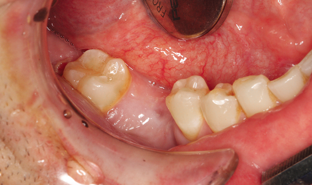
Fig. 1 An intraoral photo shows a patient missing a lower-right first molar
Imaging workflow
When it comes to planning implant surgery, my approach is to work from the crown down; that is, I plan the end and work back to the beginning. Through the use of digital imaging, I can virtually plan the entire procedure, reducing the risk of unexpected surprises during implant placement.
The List: 3 ways to maximize your intraoral camera
During my initial appointment with a patient, I will capture an image with an intraoral camera (Fig. 1).


Fig. 2 Panoramic radiograph
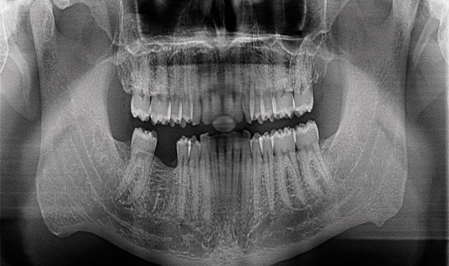
Fig. 2 Panoramic radiograph
I also take a panoramic radiograph (Fig. 2). These diagnostic tools not only aid in determining if a patient is a suitable candidate for implants, but also facilitate patient communication and case acceptance.

Fig. 3 Virtual planning implant tooth # 30, using CS 3D imaging software
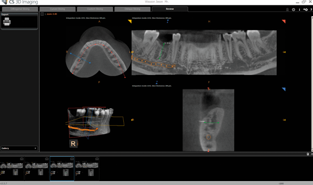
Fig. 3 Virtual planning implant tooth # 30, using CS 3D imaging software
Next, I take a 3D scan using my in-office cone beam computed tomography (CBCT) system (Carestream Dental’s CS 9300C) (Fig. 3). For as long as I can remember, having 3D information has been an important part of my implant planning and placement-I would even send my patients out for scans before I incorporated a unit into my practice. I consider 3D imaging to be the standard of care when it comes to implant procedures as it allows me to more accurately identify the morphology of the bone than 2D imaging alone.2
Must-read: Getting a clearer view of 3D imaging in your practice
Using Carestream Dental’s CS 3D Imaging software, I can virtually assess the area to determine if there is enough bone to continue or if a sinus lift, bone graft or other procedure is necessary first. If the patient is ready for an implant at that time, I can also assess the complexity of the case to determine if a surgical guide is necessary.

Fig. 4 Scan body in place ready for intraoral scan
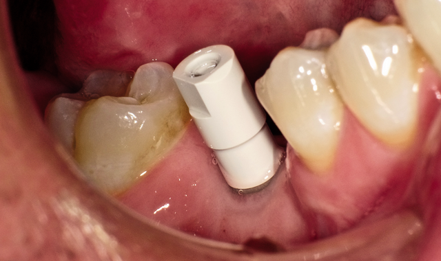
Fig. 4 Scan body in place ready for intraoral scan
Surgical guides
For cases where the CBCT scan shows there is enough bone to proceed, I can place the implant without a surgical guide by utilizing the existing CBCT with the measuring tools to decide the best implant to place virtually. If the case is more complex, I use my imaging systems for the fabrication of a surgical guide.
Approximately 80 percent of implant cases involve single teeth (Fig. 4).

Fig. 5 Screenshot image of scan progress with Carestream Dental's CS 3500
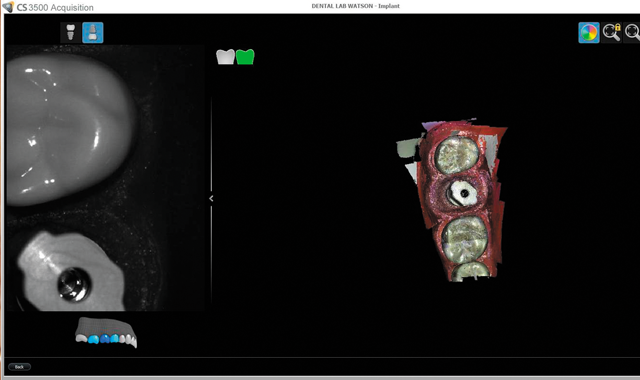
Fig. 5 Screenshot image of scan progress with Carestream Dental's CS 3500
In addition to taking the CBCT scan, I can also capture a digital impression using an intraoral scanner (Carestream Dental’s CS 3500) (Fig. 5).

Fig. 6 Surgical guide to ensure predictable prosthetic results
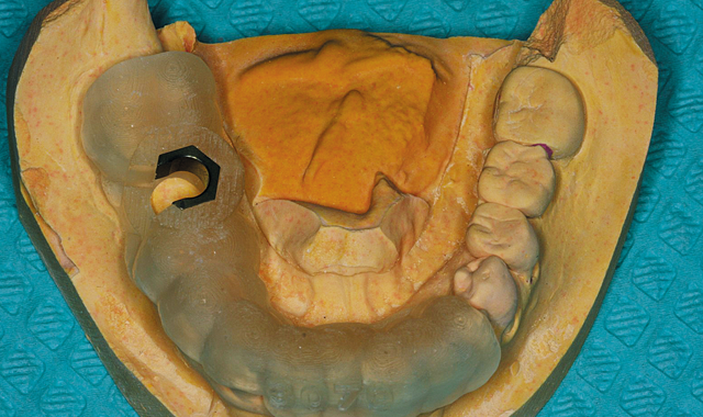
Fig. 6 Surgical guide to ensure predictable prosthetic results
From there, I can send the scans to a lab to merge the data files to make a surgical guide (Fig. 6). Both my CBCT and intraoral files are open-format (DICOM and STL, respectively), so I can send this information to the laboratory of my choice for fabrication. Merging CBCT and digital impressions is quickly streamlining dental implant surgery as well as improving accuracy of the results.3
Depending on what I see in the CBCT scan, I can order a surgical guide with pilot drills or a fully guided surgical guide that includes position, angle and depth. This is beneficial in a lot of ways, including having the ability to confidently perform the treatment plan as well as to obtain predictable results.
Must read: The hidden dangers of the gray market
The use of digital impressions-when combined with CBCT-is also important when performing full-arch dentistry. When taking out four-six teeth and placing a screw-retained provisional, imaging data allows us to see where the teeth are as well as plan implants and develop the provisional in the proper position.
Conclusion
With all of the imaging information now available to clinicians, we are better able to virtually plan treatment. The end result is more predictable surgical procedures and outcomes.
References
1. Gaviria L, Salcido JP, Guda T, Ong JL. Current trends in dental implants. Journal of the Korean Association of Oral and Maxillofacial Surgeons. 2014;40(2):50-60. doi:10.5125/jkaoms.2014.40.2.50.
2. Arora S, Lamba AK, Faraz F, Tandon S, Ahad A. Role of Cone Beam Computed Tomography in Rehabilitation of a Traumatised Deficient Maxillary Alveolar Ridge Using Symphyseal Block Graft Placement. Case Reports in Dentistry. 2013;2013:748405. doi:10.1155/2013/748405.
3. Ganz, S. The Next Evolution in CBCT: Combining Digital Technologies. Inside Dentistry. 2013; 9(2).