Finding it early
You don’t want to catch it when it’s easy to see. By then, it may be too late. As a dental professional, you need to spot possible problems in the early stages, when treatment is less invasive and chances for survival are higher.


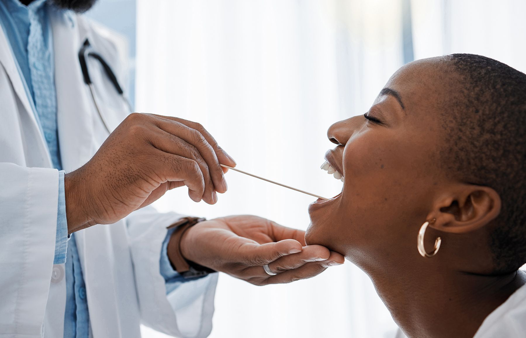
Finding It Early | Image Credit: © Kirsten Davis/peopleimages.com - stock.adobe.com
Performing a conventional white light oral cancer screening exam will help you discover potential problems and is something you should do for every patient. The exam itself only takes a few minutes and is easy to incorporate into routine appointments. But, even when you perform a thorough exam, it doesn’t mean you’ll see everything in every patient’s mouth that might be suspect. That’s where adjunctive devices come in. They can help you see what the naked eye might miss with light reflection alone, and catch subtle lesions before they become a threat.
These devices are easy to incorporate into your exam routine; you just have to do it. Depending on what adjunctive system you use, you’ll likely only add a few minutes to the appointment time. That’s not much, and patients appreciate those extra minutes-especially if you catch a problem early enough that something can be done about it.
“When I studied my own practice, I found 10 lesions that were pre-cancerous on the same patient base over one year’s time that I had not found by a conventional white light exam alone,” said Dr. Kevin Huff, who uses the VELscope in his practice of about 950 patients and published the results of his study last year. “So in my mind, direct tissue fluorescence visualization is a very effective adjunct.”
Finding the right tool for your practice
As with any technology, you need to do some research before deciding which device is best for your patients and your practice. Dr. Mark Bride, former VP, Medical Affairs for Zila Pharmaceuticals (maker of ViziLite Plus with TBlue), said the Internet is a great place to start to learn more about these discovery aids and what they offer.
Peer-reviewed articles can be found on the Internet and provide a wealth of information. Talking with manufacturers and actually seeing the products in use at tradeshows also is a great way to compare the different options and determine which one will best fit into your practice. Beyond that, there are some good online courses and dental lectures available that thoroughly cover the pros and cons of all of the adjunctive screening devices.
Every practice is different, so you have to choose a device that you know you and your team members can easily incorporate into the patient exam, said Dr. Andres Zuluaga, VP of R&D at Trimira, the company that makes the Identafi 3000 ultra. If you can’t get out to a show, contact the manufacturer directly and ask for an in-office visit. Make sure you feel comfortable with the manufacturer and that the company offers the necessary training to get you and your staff up to snuff on using your new discovery tool.
Once you make the decision to purchase one of these devices, you next have to determine how you’re going to charge for it, or even if you are going to charge for it, Dr. Huff said. Some doctors charge a flat fee that could range from $40 to $200, while others raise the price of the exam $4 or $5 to cover costs rather than charging an additional fee. The route you go likely will depend on the tool you choose and where your office is located, but if you want patients to agree to the extra screening you have to keep it affordable.
Making sure you use it
It’s easy to buy one of these devices with every intention of using it only for it to end up collecting dust somewhere in the back of the office. But that doesn’t do your patients or your practice any good.
Incorporating these adjunctive exams into your routine is easy. For ViziLite Plus, making sure you put it on the tray before the patient even gets into the office acts as a reminder to do the exam, Dr. Bride said. Don’t look for reasons why you can’t use it.
“It’s just changing the mentality of the practice that there’s one additional step to do,” Dr. Bride said. “And the time it takes (about 90 seconds) isn’t an excuse not to do it.”
Dr. Huff uses his VELscope for every new patient, and his hygienist uses it during every recall visit, working it in to the exam routine immediately after the conventional oral exam. If you know you’re doing the exam regardless of risk factors, which are changing, you and your team members won’t forget it needs to be done. You’ll just do it because it’s part of the normal exam routine. And with HPV 16 now a known cause of oropharyngeal cancer, you can’t limit your screenings to your older patients who smoke and/or drink. Your younger patients are at risk now, too.
Talking to patients about it
While you’ll have some patients who come to your office specifically because they know you have one of these devices, there are others who have no idea what a VELscope or ViziLite does or why they should care. This is where patient education comes in.
Patient brochures in the reception area, educational videos and even posters are great ways to educate your patients about oral cancer and adjunctive devices like the Identafi 3000 ultra, said Jerry Trzeciak, VP of Sales and Marketing at Trimira. When they’re in your chair ready for the screening, take the time to explain to them what you’re doing and why. Let them know this is just a screening aid that helps you see what you can’t see with a conventional exam alone, and that it’s painless and something you offer every patient.
“Educate patients about the disease and about the technology so they look at it as a real opportunity for early detection as opposed to fearing something,” Trzeciak said. “Have your staff use the technology prior to offering it to patients so they can experience the technology themselves. They can then share that experience with confidence and say it is uninvasive, easy to use and patient friendly.”
What to do if you find something
If you find something suspicious during one of these exams, do exactly what you would do if you found it during the conventional screening. First, try to figure out what the cause could be. If you find an ulcer or a lesion on the roof of a patient’s mouth, ask the patient if there’s anything that could have caused the trauma and if he or she knows how long it’s been there. Ask questions and look around the mouth to determine the source and eliminate the possibility that it’s a sign of cancer.
It’s also important to photo document your findings, which is something Dr. Huff said is easy to do with the VELscope with a simple camera attachment and a point-and-shoot camera. This also is the time a suspicious lesion can be marked with Zila’s TBlue, Dr. Bride said.
“No matter what you do, try to avoid scaring your patients. If you find something that’s suspect, avoid using the word cancer,” Dr. Huff said. “Take a closer look at the area and if you can’t rule cancer out, tell the patient you found something that’s irregular or unusual and you’d like to see him or her back in two weeks.”
Making the referral
If the suspicious lesion is still there when the patient returns in two weeks, it’s time for a referral. Sometimes cytology with a brush can be helpful as a tool for patient education or to strengthen your reason for a surgical biopsy, Dr. Huff said.
Remember, making a diagnosis is not your job as a general dentist, but recognizing something abnormal is. Unless you are comfortable performing biopsies yourself, you need to refer the patient to an oral surgeon for biopsy. The tissue sample should then be sent to an oral pathologist to find out exactly what you’re dealing with, said Brian Hill, Executive Director of the Oral Cancer Foundation.
There are a lot of things that can go wrong in a person’s mouth, and while you’re not expected to learn everything about every lesion out there, it is your job to discover abnormalities and refer the patient to someone who can make a diagnosis. You don’t have to know what it is you’re looking at, but you do need to recognize abnormalities. Is it a different color than surrounding tissue? Does it bleed easily when you touch it? Is it hard or raised above the surrounding tissue? All of these are characteristics of lesions that need to be biopsied if they persist.
“Nobody is going to laugh at you if you refer something out that is benign. You need to refer all suspicious and persistent abnormalities,” said Hill, who also is a stage 4 oral cancer survivor. “As a dental patient, I would much rather someone biopsy something they thought was suspect, and later say it turned out to be nothing, than guess, or watch and wait. A biopsy would yield a gold-standard, black-and-white answer. I would have been anxious while waiting for a couple days to get the pathology report, but to hear that it’s nothing serious is a golden moment. That’s better than having someone tell you it doesn’t look like oral cancer, and then do nothing to find out for sure.”
Getting staff involved
No matter which tool you decide to go with, your staff has to be on board, Dr. Bride said. Depending on the statues of each state’s Dental Board, the majority of the time a staff member executes the exam and then alerts the doctor if there’s something suspicious. That’s why team training and education is so important, Trzeciak said. The staff should be educated about the disease state, the risk factors and what can happen if a patient is diagnosed with this disease in a late stage. They have to know how to use the screening tool and why it’s important.
Why early detection matters
Patients diagnosed with oral cancer often have to undergo severe treatments to survive. If they do survive, they often have to go through life without their voice, or the ability to eat solid foods. The realities of this disease can be harsh, and the earlier it can be detected the better the chance of survival and minimal treatment-related complications.
And with news of celebrities like Michael Douglas battling this disease, more and more of your patients are becoming aware of oral cancer and what it can do. They’re asking for thorough screenings and often are appreciative when a clinician or hygienist takes the time to perform an extra exam that may end up saving their life.
Whichever adjunct you choose, incorporating it into practice is easy. Make a spot for it after the conventional exam. You’ll soon discover you’re finding lesions you never would have seen before. While most of these won’t be cancer, the extra effort will be well worth it for the ones that are.
“Dentists are so much more than drill, fill and bill kind of people. As custodians of their patients’ oral health, they should be involved in everything that goes on in a patient’s mouth,” Hill said. “Oral cancer is only one. But it’s probably the only one that’s going to take a life.”


