An up close look at an innovative caries detection device from DEXIS
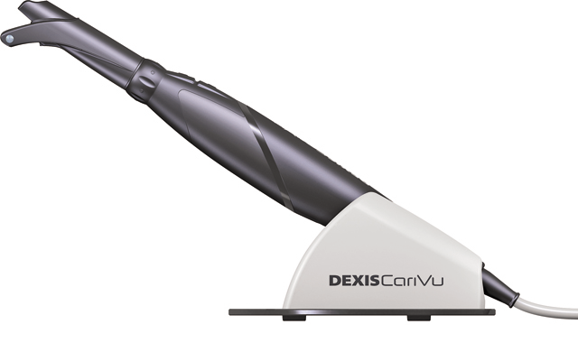
CariVu from DEXIS offers the clinician a precise picture of caries by using near infrared transillumination technology to give clinicians a view of carious lesions and cracks.

The percentage of dentists switching from film-based X-ray to digital radiography continues to grow. One of the many benefits of digital imaging systems is reduced radiation. The American Dental Association supports this, citing that clinicians can lower radiation exposure to their patients between 40-60 percent, depending upon the system.
DEXIS Platinum sensors and software already provide consistent image quality at the lowest radiation dose†, and with the addition of DEXshield™, designed exclusively for use with the DEXIS™ Platinum sensor, this exposure is reduced even more -- by an additional 30 percent. Even with all the efforts that are happening to reduce radiation doses, there are still patients whose reluctance to have X-rays or whose medical history preclude the use of any type of radiography.
For caries detection, DEXIS CariVu™ is an innovative and useful device that emits absolutely no ionizing radiation. For all patients, whether they are anxious about radiation or not, what is better than low dose? No dose!
Related reading: Dr. John Graeber discusses how CariVu has impacted his dental practice and patients
Detection with a difference

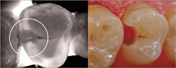
CariVu offers the clinician a precise picture of caries by using near infrared transillumination technology to give clinicians a view of carious lesions and cracks. Flexible “arms” on the end of the device tip straddle the tooth, and near infrared photons are emitted through the arms and travel from the root to the crown of the tooth. Dense enamel reflects the photons while porous lesions trap and absorb the photons.
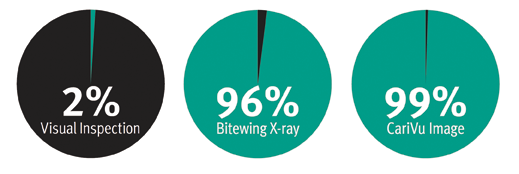
As a result, healthy tooth appears light, and lesions appear dark, giving clinicians a structural view of the lesion whether it’s interproximal occlusal, or around restorations or a crack. As the old saying goes, “What you see is what you get,” or as DEXIS likes to say, “What you see is what is there.” CariVu technology has a very high detection accuracy -- an interproximal dentin caries detection rate of 99 percent. As a result, clinicians can obtain highly accurate diagnostic images in a noninvasive manner with absolutely no ionizing radiation.
For more on CariVu: DPR tech editor gives thumbs up to the DEXIS CariVu transilluminator

CariVu’s transillumination technology is different than other caries detection devices, like fluorescence imagers or scanners that deliver an “indication” of caries, not the precise picture. Fluorescence imagers bathe teeth in blue-purple light most often used for soft tissue screening. Scanners aim yellow-red light directly onto the area. These units present an image with a range of colors, or they display a number correlated to a degree of demineralization. This type of detection has limitations. Bacteria, restorative materials and calculus can yield false readings, and some scanners can only be used on occlusal surfaces.
Those in-between times

Using X-ray technology with a CariVu system can give the clinician a more comprehensive view of the development of caries. CariVu is a great asset during recall visits, especially when patients are not due for bitewings. Since there is always a chance that decay may spread before X-rays are indicated, capturing a quick CariVu image of the area can let dentists know if it’s appropriate to wait to take radiographs until the next recall visit, or if they should begin preventive or restorative treatment right away.
CariVu is also helpful for monitoring suspicious areas between routine exams. If a questionable area is identified on a radiograph – especially interproximal decay, which can be more difficult to detect -- the “second opinion” can be a transilluminated image that reveals the extent of the condition and helps to confidently determine whether it needs monitoring over time or requires immediate treatment.

Compatible with trusted DEXIS software
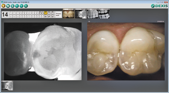
In addition to its innovative technology, CariVu is fully integrated with the DEXIS imaging software programs. Dentists are able to display the transillumination video feed or images next to previously captured X-rays and camera images, allowing for quick comparison and monitoring of areas over time. Since CariVu images are similar in appearance to familiar X-ray images, there is no need to calibrate the device or become versed in the meaning of multiple color codes or numeric indicators to find out the location and size of caries.
More on CariVu
CariVu is a compact, portable caries detection system that features patented transillumination technology. Designed to incorporate easily into current workflows, CariVu lets dental clinicians easily detect a range of carious lesions and cracks, plus it provides an easy-to-interpret image that is stored with the patient’s additional images. For more information, contact DEXIS at 888-883-3947 or dexis.com.