Advertorial: A New 3D Vision for Dentistry
Take a Look at Stationary Intraoral Tomosynthesis, a 3D Radiographic Imaging Technology

Visibility is a key ASPECT to successful dental treatment, from external lighting and magnification to intraoral and extraoral imaging. When a dental technology offers new approaches to visualizing the oral environment it’s worth taking note. This is certainly the case with stationary intraoral tomosynthesis (sIOT).
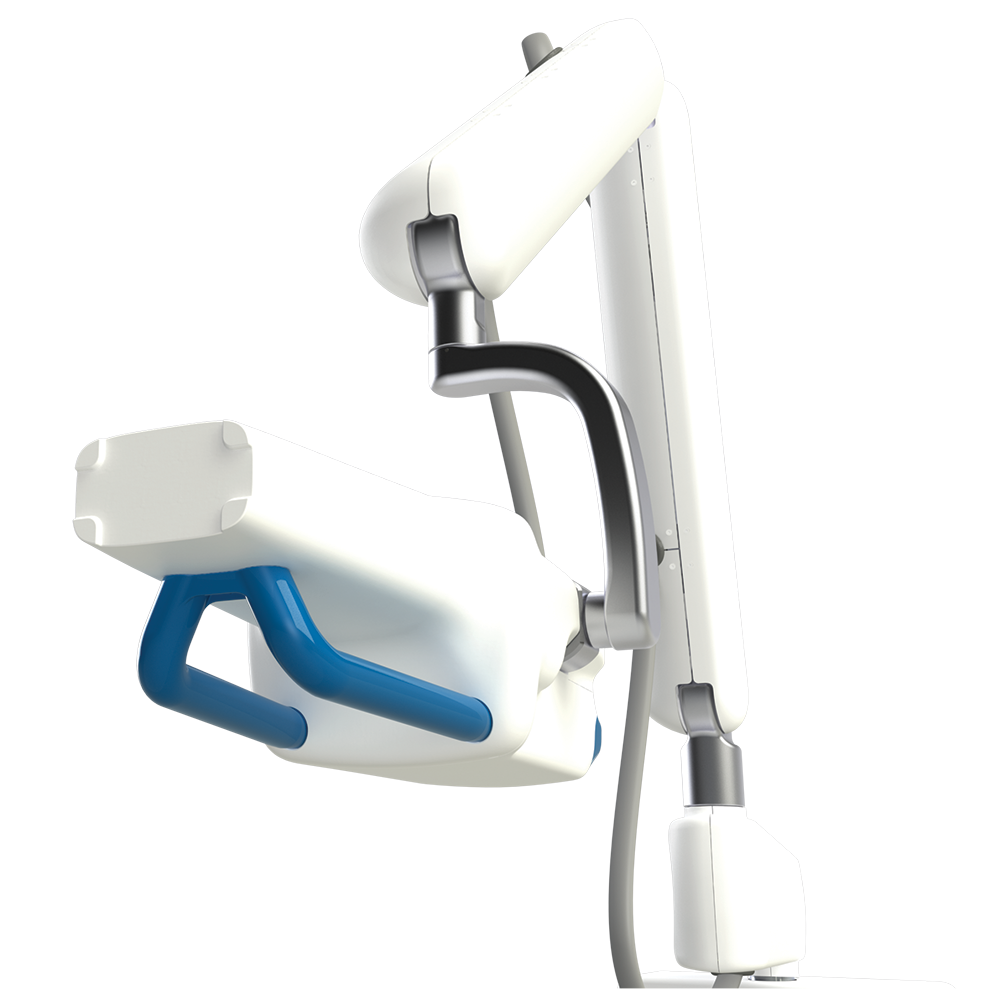
A technique for producing 3D radiographic images intraorally, sIOT captures multiple x-ray images simultaneously via carbon nanotube x-ray sources and then processes those images using algebraic reconstruction to produce a stack of 2D tomographic x-ray layers each 70 to 80 µm thick. The technology has the potential to enhance clinical diagnostics and improve other aspects of dental care, says Donald Tyndall, DDS, PhD, MSPH, FICD, a professor in the Division of Diagnostic Sciences at University of North Carolina at Chapel Hill Adams School of Dentistry, who works on developing sIOT technology for dental use.
Dental Imaging Frontiers
When discussing the promise of sIOT, Dr Tyndall starts with the history of dental imaging. The first intraoral x-ray image was captured in 1896, and although it is much faster and image quality is vastly improved today, the process is essentially the same. A generator shoots x-rays at a receptor and an images of the insides of any structures in between are captured.

Traditional dental intraoral and panoramic x-rays face the same challenge of trying to capture 3D structures in a 2D format. “In a way, we’re still imaging like it’s 1896,” he says. “We’re collapsing a 3-dimensional object into 2 dimensions, and this leads to issues of overlap. We’re somewhat limited by not so much the technology, but by geometry.”
Dr Tyndall says 2D imaging reamains useful, but 3D imaging truly takes dentistry to a different level when it comes to diagnostics, treatment planning, case presentation, and patient education.
“There are data showing that patients are more likely to accept a treatment plan if they see it in 3D,” he says. “The more the patient understands, the more likely they are to accept the treatment plan.”
Cone beam imaging has made a huge impact on dental diagnostics and treatment planning, but Dr Tyndall notes this 3D technology has limited value in caries detection and there are challenges in imaging around metal artifacts. With sIOT, dentists now have a 3D imaging approach that can address those issues.
sIOT Explained
Images captured with sIOT technology look similar to a traditional dental x-ray, but because they are digital composites of multiple images, they allow a clinician to look through the depth of a tooth 1 layer at a time instead of viewing a single image comprised of that same information collapsed into a single plane. A sIOT system uses an intraoral sensor and a custom x-ray generator designed to work together to capture multiple images at once.
“We’re staying in the same plane but we’re moving the x-ray source to different locations, and so we’re generating multiple pictures,” Tyndall explains.
Similar to how humans gain depth perception by processing images from 2 eyes set a slight distance apart, sIOT systems create x-rays with depth by constructing an image based on multiple images captured from slightly different distances and angles.
The images can be captured using the same imaging workflow for intraoral x-rays and they can be read with little additional training because they look like traditional 2D x-rays. Additionally, because the tuned aperture CT technology used in the image reconstruction is able to avoid metal artifact interference, sIOT images do not face challenges when imaging implants, orthodontic appliances, and other metal objects.
sIOT does not produce a 3D image that can be manipulated on screen like a CBCT image. Instead it the viewer can move backward and forward through the layers on a plane parallel to the x-ray sensor.
Because sIOT imaging doesn’t superimpose structures on top of each other, the images make it easier to spot defects such as cracks and lesions. Dr Tyndall says this is especially true when it comes to proximal areas because, whereas traditional x-rays often show closed contacts due to overlap, at least 1 layer in a sIOT image is likely to open that contact, reducing the need to retake images. Other imaging advantages include greater visibility of the periodontal ligament and the ability to view buccal and palatal roots separately when examining molars.
Although the sIOT technology provides a lot more information than a traditional dental x-ray, the imaging requires more time and a slight increase in radiation exposure for the patient. Still, Dr Tyndall notes that radiation levels currently required for sIOT imaging are not putting patients at much greater risk than they face from other dental radiograph technologies.
“Think of it as intraoral imaging with slightly more dose, but that slightly more dose means a lot more information,” he says.
Applications for sIOT
Although the technology is relatively new, sIOT is already approved for clinical use in the US. In August 2021 the US Food and Drug Administration announced 510(k) premarket clearance for Surround Medical Systems’ PORTRAY imaging system, making it the first sIOT solution designed for dental use.
The technology can be used for standard radiographic imaging such as bitewings or a full mouth series. In those cases it can provide more information on each tooth than can be provided by a standard x-ray series. Dr Tyndall believes sIOT is a great option for any alveolar imaging, as well as any case where a bitewing or periapical x-ray would be of use. These images can be advantageous for caries detection, periodontal bone assessment, implant site assessment, fracture detection, and more. The fact that the images will almost always capture open contacts can also mean fewer retakes.
“Stationery intraoral tomosynthesis might be another great way of gaining extra 3D information about the teeth and periodontia,” Dr Tyndall says.
The Future
Dr Tyndall says he is excited about the future of dentistry with sIOT, and he plans to continue to study the technology’s applications. One thing he sees in the future of sIOT is artificial intelligence integration to make enhance the efficiency of analyzing the information captured by system.
Another technology Dr Tyndall is watching is dental MRI, a new, radiation-free approach to intraoral imaging that is distinct from medical MRI. Although he believes these systems are several years away, he believes dental MRI will be useful for imaging cysts and tumors, as well as inflammation and nerves.
With sIOT already here, and other 3D imaging technologies on the way, Dr Tyndall is excited about the future of dental care. He’s confident that enhanced and varied 3D imaging technologies, as well as other 3D digital dental solutions, will improve detection and diagnostics, enhance treatment planning and case presentation, and help heighten the quality and efficiency of dental care.
“3D won’t be the diving board into the future of radiographic imaging and dentistry. It’ll be in the pool itself,” he says.