How to navigate the new periodontal classification system
The new proceedings may appear confusing, but becoming familiar with them will lead to skillful dental hygiene decision-making.


The proceedings of the 2017 World Workshop on the Classification of Periodontal and Peri-Implant Diseases and Conditions was published in June 2018 and it’s a revision of a 1999 World Workshop Classification. The new proceedings are a bit of a maze to navigate because they contain multiple case definition papers and consensus reports. The papers and consensus reports are a joint effort by the American Academy of Periodontology and the European Federation of Periodontology. The intent of the workshop was to base classification on strongest scientific evidence when available; otherwise, lower levels of evidence and expert opinion were used.1
Changes in gingivitis/periodontitis classification
I’m not convinced that many clinicians are familiar with the 1999 periodontal disease classifications, but I remember lecturing on it to my students and writing about it. From 1999- 2017, periodontitis was classified as chronic (localized and generalized), aggressive (localized and generalized), necrotizing and as a manifestation of systemic disease. Gingivitis was differentiated as dental plaque-induced and non-plaque-induced.2 The newly published classification system (2018) includes characteristics of peri-implant tissues in health and disease that weren’t included in the 1999 classifications.3
Three forms of periodontitis have been agreed upon and identified:
1. Necrotizing periodontitis
2. Periodontitis as a manifestation of systemic disease
3. Periodontitis
Forms of disease previously identified as chronic or aggressive have been removed and are now grouped under the single category “periodontitis.”1 The workshop agreed on a classification framework for periodontitis based on a staging and grading system that can be adapted over time to reflect new scientific evidence.1
Staging means determining the severity of disease at presentation and the complexity of disease management.
- Stage 1: Initial periodontitis
- Stage 2: Moderate periodontitis
- Stage 3: Severe periodontitis with potential for additional tooth loss
- Stage 4: Severe periodontitis with potential for loss of dentition
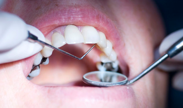
Extent and distribution of disease should be noted: localized, generalized and molar-incisor distribution. Staging isn’t something that can be determined by recording probing depths only and takes time and critical thinking skills to assess. Several variables need to be carefully evaluated, including clinical attachment loss (CAL), amount and percentage of bone loss, probing depth, presence, and extent of angular bony defects and furcation involvement, tooth mobility and tooth loss due to periodontitis.
Grading includes three levels:
- Grade A: Slow rate of progression
- Grade B: Moderate rate of progression
- Grade C: Rapid rate of progression
When grading a patient, general health status and other exposures like smoking or level of metabolic control in diabetes need to be taken into account to allow comprehensive patient management.
Take, for example, Gena Gentry, age 32, who presents as a new patient. She weighs 230 pounds and was recently diagnosed with Type 2 diabetes, hypertension and hyperlipidemia. In questioning her about her HbA1c, she reports A1c > 9 percent as of last week. In examining her periodontium, her assessment is summarized as: Periodontitis associated with diabetes mellitus, Stage II, Grade C. In staging Gena, CAL, radiographic bone loss and probing depths were taken into consideration. Grading severity was based on smoking, obesity and HbA1c.4 In order to stage and grade your patient, you’ll need a copy of the staging and grading framework. You can print it out for free at www.perio.org. (Click on 2017 World Workshop Proceedings, click again on the Journal of Periodontology Proceedings, and then scroll down until you find the document by Tonetti, Greenwell and Kornman titled “Staging and Grading of Periodontitis.”) Read the document, laminate it and share it with your professional colleagues.
In 1999, gingivitis was referred to as gingival diseases and categorized as dental plaque-induced gingival diseases or non-plaque-induced gingival diseases. The newly published classification system differentiates between plaque-induced gingivitis and non-plaque-induced gingival diseases. Management of gingival inflammation is a key risk factor for the onset of periodontitis, and plaque-induced gingivitis results from supra- and subgingival plaque/biofilm accumulation modified by many factors, including systemic diseases/disorders like diabetes and oral factors such as restorative margins, xerostomia, drug-induced gingival enlargement, and tooth and root anatomy.
Non-plaque-induced gingival diseases are less often diagnosed but should be reviewed periodically. Lichen planus, traumatic lesions and necrotizing stomatitis are some common presentations. Non-plaque-induced gingival diseases can be reviewed at www.perio.org with free access to 2017 World Workshop documents pertaining to gingival diseases and gingivitis.
The 2017 World Workshop Proceedings introduced a new term for plaque-induced gingivitis on a reduced periodontium. A reduced peridontium results following active periodontal treatment and the resolution of periodontitis-related inflammation. The periodontal tissue is clinically non-inflamed with a reduced connective tissue attachment and alveolar bone height. Plaque-induced gingivitis on a reduced periodontium has the following characteristics:
- No evidence of progressive attachment loss (active disease)
- Pre-existing attachment loss with a higher risk of professional supportive maintenance care.
Staging of plaque-induced gingivitis is recommended as follows:
- Localized gingivitis: BOP score ≥ 10 percent and ≤30 percent, without attachment loss and radiographic bone loss.
- Generalized gingivitis: BOP score > 30 percent, without attachment loss and radiographic bone loss.
Gingivitis on a reduced periodontium can be graded as localized (BOP ≥ 10 percent and ≤ 30 percent) or generalized (BOP > 30 percent). Examples of gingivitis patients with a reduced periodontium are those with gingival recession and crown lengthening who don’t have a history of periodontitis.
Peri-implant case definitions
The 2017 World Workshop case definitions for periodontal lesions around dental implants focus on biofilm-induced inflammatory lesions only. As we already know, dental implants and their restorations are subject to mechanical and biological considerations that can affect long-term prognosis.6
World Workshop participants defined the following terms: peri-implant health, peri-implant mucositis and peri-implantitis. The case definitions are based on a systematic review of the scientific evidence and correlates with clinical and radiographic findings for the three entities.6 The case definitions proposed by the 2017 World Workshop participants are only intended to apply to biofilm-induced inflammatory lesions.
Peri-implant health requires:
- Visual inspection to rule out signs of inflammation: pink as opposed to red and swollen tissue. Consistency of tissue should be firm and not spongy.
- Lack of profuse or any other bleeding on probing.
- A significant increase in probing depths over time. Keep in mind, however, that probing depths can differ, depending on the height of the soft tissue at the implant location.
- Absence of further bone loss following initial healing, which should not be ≥ 2 mm.
Peri-implant mucositis
The American Academy of Periodontology has defined peri-implant mucositis as a disease that includes inflammation of the soft tissues around a dental implant without additional bone loss after the initial bone remodeling that may have occurred during healing after the surgical placement of the implant.6 Case definition of peri-implant mucositis includes:
- Local swelling, redness and shininess of the soft tissue, which are classic signs of inflammation.
- A local dot of bleeding that isn’t the result of traumatic probing.
- Any bleeding on probing that’s accompanied by visual inflammatory changes.
- Suppuration upon clinical examination (following probing or gentle pressure to the tissues).
"Intraoral radiographic evaluation of bone levels around implants should always be included in the presence of clinical signs of inflammation. In addition, a prerequisite for the evaluation is that a radiograph be taken at baseline (suprastructure in place) and used for future assessment of mesial and distal bone levels in relation to defined references. Accounting for the remodeling process of alveolar bone during the first year after installation, the change in bone level since the placement of the prosthetic suprastructure should not be > 2 mm. Presence of bone loss beyond crestal bone level changes resulting from the initial remodeling process of alveolar bone after implant installation suggests either progressive peri-implant infection or other local factors such as excess cement and looseness/fracture of implant components.”6
Peri-implantitis
The progressive loss of supporting peri-implant bone distinguishes peri-implantitis from peri-implant mucositis. Peri-implantitis is an inflammatory lesion of the mucosa surrounding an endosseous implant that shows evidence of any additional bone loss following the initial implant installation and loading. During initial implant placement, some crestal bone height is lost during the healing process and can range from 0.5-2 mm.6
The diagnosis of peri-implantitis includes:
- Visual inflammatory changes in peri-implant tissues combined with bleeding on probing and/or suppuration.
- Increasing probing depths as compared to measurements obtained at placement of the supra-structure.
- Progressive bone loss in relation to the radiographic bone level assessment at one year following delivery of implant-supported prosthetics reconstruction.
- In the absence of initial radiographs and probing depths, radiographic evidence of bone level ≥ 3 mm and /or probing depths ≥ 6 mm in conjunction with profuse bleeding.
The 2017 World Workshop Classification of Periodontal and Peri-Implant Diseases and Conditions also includes literature on periodontal abscesses, necrotizing periodontal diseases and endo-periodontal lesions and they’re grouped together according to common features. Dental practitioners need to review this chapter and another longer one on systemic conditions that have a major effect on the course of periodontitis like smoking and diabetes. A novel classification of traumatic occlusion lesions is also discussed and the importance of biological width (now called supracrestal tissue attachment) is emphasized and discussed. Visit www.perio.org and follow the aforementioned instructions to get free access to these additional documents.
Spend time studying and applying the newly revised perio classification system for everyday assessment of new patients and patients of record. If you’re really serious about this topic, print out and laminate case definitions that you need to reference as you examine a patient. Periodontal case assessment requires critical assessment skills and it’s an essential process for safe, efficient and skillful dental hygiene decision-making.
References:
1. Caton JG. A new classification scheme for periodontal and peri-implant diseases and conditions- introductions and key changes from the 1999 classification. J Periodontol. 2018; 89 (Suppl 1):S1–S8.
2. Armitage GC. Development of a classification system for periodontal diseases and conditions. Ann Periodontol. 1999; 4(1): 1-6.
3. Stefan RG et al. Peri-implant health, peri-implant mucositis, and peri-implantitis: Case definitions and diagnostic considerations, J Periodontol 2018 ; 89, S1, (S304-S312).
4. Tonetti MS, Greenwell H, Kornman KS. Staging and grading of periodontitis: framework and proposal of a new classification and case definition. Periodontol. 2018;89(Suppl 1):S159–S172.
5. Murakami S et al. Dental plaque-induced gingival conditions. J Periodontol. 2018;89(Suppl 1):S17–S27.
6. Renvert S et al. Peri-implant health, peri-implant mucositis, and peri-implantitis: case definitions and disgnostic considerations. J Periodontol. 2018;89(Suppl 1):S304–S312.


